Skin lesions is a broad term for various visible or palpable changes in the skin: patches, nodules, raised areas, wart-like growths, pigmented areas, vascular lesions, rough patches, subcutaneous lumps, scar-like changes or non-healing sores. Some skin lesions are benign and pose no threat to health, while others may be precancerous changes or skin cancer.
Most importantly: appearance alone is often not enough for a person to determine reliably what a skin lesion is. A lesion should be checked by a dermatologist if it is new and continues to change, grows rapidly, bleeds, ulcerates, does not heal, changes colour or shape, becomes painful or looks different from the others.
What are skin lesions?
A skin lesion is not one specific diagnosis. It is a descriptive term used for a visible or palpable change in the skin. It may be a mole, wart, skin tag, seborrhoeic keratosis, angioma, dermatofibroma, lipoma, cyst, actinic keratosis, inflammatory nodule, scar or skin tumour.
Some lesions are pigmented, such as moles. Others are skin-coloured, rough or wart-like. Soft, skin-coloured growths in skin folds are often called papillomas in everyday language. Round or mobile lumps felt under the skin may sometimes be cysts, lipomas or other subcutaneous lesions.
Not every raised skin change is a tumour. Small, solid raised areas are called papules in dermatology, and they may occur as part of acne, rosacea, psoriasis, dermatitis, an infection or another skin condition. There are also temporary skin reactions, such as urticaria or hives, which may look like raised, itchy lesions but are usually not persistent skin growths.
Why is “skin lesion” not a single diagnosis?
Lesions that look alike may arise from different skin structures: pigment cells, epidermal cells, blood vessels, connective tissue, fat, hair follicles, scar tissue or inflammatory processes. Their treatment and risk assessment therefore differ.
For example, a raised brown lesion may be a mole, seborrhoeic keratosis or a pigmented skin tumour. A rough patch may be dry skin, a wart, actinic keratosis, psoriasis or an early form of squamous cell carcinoma. A subcutaneous lump may be a cyst, lipoma, inflamed follicle or another lesion. A thickened raised area after injury, surgery or inflammation may be a hypertrophic scar or keloid. This is why examination by a dermatologist and, when appropriate, dermoscopy are important.
Common types of skin lesions
The table summarises common skin changes. These descriptions are for guidance and are not sufficient for self-diagnosis: benign and malignant lesions may look similar.
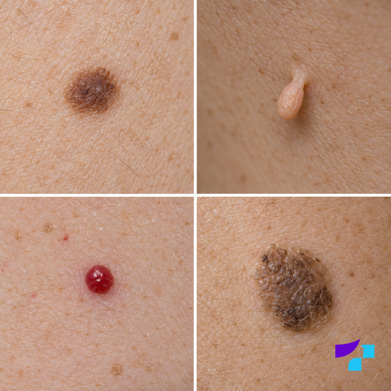
The image shows examples of common benign skin lesions: a mole, a skin tag (acrochordon), a cherry angioma and a seborrhoeic keratosis. The image is illustrative; a diagnosis cannot be made from appearance alone.
| Type of lesion | What it may look like | Usually benign? | When should it be checked? |
|---|---|---|---|
| Mole | A brown, black, bluish, skin-coloured or raised lesion | Usually | If it changes, bleeds, becomes asymmetrical or looks different from the others |
| Wart | A rough, firm raised lesion, often on the hands or feet | Usually | If it hurts, grows, bleeds, is on the face, near a nail, on the foot or genitals, especially if the diagnosis is unclear |
| Papilloma or skin tag | A soft, skin-coloured or brownish growth, often on a stalk | Usually | If it is repeatedly injured, bleeds, grows, becomes inflamed or the diagnosis is unclear |
| Seborrhoeic keratosis | A waxy, rough, brown or greyish lesion that may look “stuck on” | Yes | If it looks atypical, changes rapidly or resembles melanoma |
| Actinic keratosis | A rough, scaly patch on sun-damaged skin | A precancerous change | Assessment by a dermatologist is advisable |
| Angioma | A bright red or purplish-red dot or small nodule | Usually | If it bleeds, changes rapidly, becomes dark or causes problems |
| Haemangioma | A red or bluish vascular lesion; depending on the type, it may be visible at birth or appear during the first weeks of life | Yes | If it grows rapidly, ulcerates, bleeds, affects function or is in a sensitive location |
| Dermatofibroma | A small, firm, brownish or pink nodule, often on the legs | Yes | If it grows, hurts or the diagnosis is unclear |
| Cyst | A round subcutaneous lump, sometimes mobile or inflamed | Usually | If it grows, hurts, becomes inflamed, discharges material or recurs |
| Lipoma | A soft, mobile subcutaneous lump | Yes | If it grows rapidly, hurts, lies deep, is difficult to move or the diagnosis is unclear |
| Scar or keloid | A flat, depressed or raised change after injury, surgery, acne or inflammation | Not a malignant lesion | If it grows, itches, hurts, bleeds, ulcerates or there is no clear preceding injury |
| Neurofibroma | A soft, skin-coloured or slightly brownish lesion | Usually | If there are multiple lesions, growth, pain or other symptoms |
| Sebaceous hyperplasia | Small yellowish or skin-coloured bumps, often on the face | Yes | If the lesion resembles basal cell carcinoma or the diagnosis is unclear |
| Papules | Small, solid bumps or nodules in the skin | Depends on the cause; a papule describes a type of change, not a diagnosis | If they persist, spread, hurt, itch or leave scars |
| Skin cancer | A non-healing sore, nodule, recurring crust or changing patch | No | It should be checked as soon as possible |
Benign, precancerous and malignant lesions
Benign skin lesions
Benign skin lesions often grow slowly, are relatively symmetrical, remain unchanged for a long time and do not bleed without injury. They may include many moles, seborrhoeic keratoses, skin tags, angiomas, dermatofibromas, lipomas and cysts. Scars are not malignant tumours either, although they may cause pain, itching or functional problems. These features alone cannot rule out skin cancer with complete certainty without medical assessment.
Precancerous changes
Precancerous changes are not yet invasive cancer, but they contain cellular changes that may progress in some cases. A typical example is actinic keratosis, which most often develops on sun-damaged areas such as the face, ears, scalp, backs of the hands and forearms.
Malignant skin lesions
Malignant lesions include melanoma, basal cell carcinoma and squamous cell carcinoma. They may be pigmented, pink or skin-coloured. Read the article on skin cancer for a broader overview of these diagnoses.
Moles and pigmented lesions
A mole, or melanocytic naevus, is a lesion formed by pigment-producing cells. It may be present at birth or appear during life. Many moles are benign, but special attention should be paid to moles that are new, changing or different from the others.
The ABCDE rule
The ABCDE rule can help with self-examination:
- A — asymmetry: one half of the lesion differs from the other.
- B — border: the edges are irregular, notched or poorly defined.
- C — colour: the lesion contains several colours or uneven pigmentation.
- D — diameter: attention should be paid to lesions larger than about 6 mm, although melanoma can also be smaller.
- E — evolving: the lesion changes in size, shape, colour or sensation over time.
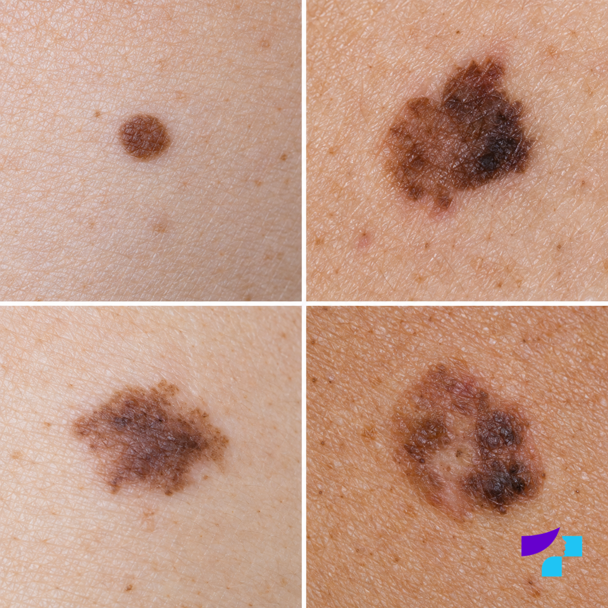
The upper-left panel shows a regular pigmented lesion, while the other panels illustrate asymmetry, irregular borders and uneven pigmentation. These features are not a diagnosis in themselves, but they are reasons to have a lesion examined by a dermatologist.
The “ugly duckling” sign is also important: a lesion that looks markedly different from the others should be shown to a doctor. The ABCDE rule does not cover every form of melanoma, so a new, growing or bleeding lesion also requires assessment even if it does not match these features. Read more in the articles on moles and mole checks.
Warts, papillomas and virus-related lesions
Warts
Warts are lesions of the epidermis caused by the human papillomavirus, or HPV. They are often rough and firm and may occur on the hands, feet, around the nails or elsewhere. Not every rough lesion is a wart: a corn, seborrhoeic keratosis, mole or even a skin tumour may look similar.
Papillomas and skin tags
In everyday language, papillomas often means skin tags or acrochordons: soft, skin-coloured or brownish growths commonly found on the neck, in the armpits, beneath the breasts, in the groin or around the eyelids. They may cause cosmetic concern, rub against clothing or jewellery and occasionally become inflamed after injury.
A skin tag is not the same as a virus-related wart. Its presence alone does not indicate HPV infection or a sexually transmitted infection. A name alone is not enough to establish the diagnosis because different stalked or wart-like growths may look similar.
A lesion that hurts, bleeds, grows rapidly or is located on the face, near a nail, on the foot or in the genital area needs assessment by a dermatologist. Read about treatment in the articles on wart removal and papilloma removal. Genital warts and HPV infection are a separate subject covered in greater detail in the article on the human papillomavirus.
Cysts and subcutaneous lumps
Subcutaneous lumps are not always skin tumours in the usual sense. They may be cysts, lipomas, inflamed hair follicles, enlarged lymph nodes or other lesions. A cyst is generally an enclosed structure with a wall; depending on its type, the cavity may contain fluid, keratin or other material. An epidermoid cyst, in which keratin accumulates, is a common type of skin cyst.
A cyst may remain quiet and painless, but it can become inflamed, red, painful, larger or discharge material. A subcutaneous lump should be shown to a doctor if it grows, repeatedly becomes inflamed, hurts, changes or its nature is unclear. A deep, rapidly growing or fixed lump may require additional investigation, such as ultrasound.
Red skin lesions and vascular changes
A red skin lesion does not always indicate inflammation or cancer. It is often a vascular lesion, such as an angioma or haemangioma. Cherry angiomas usually look like small, bright red or purplish-red dots or bumps. They occur more frequently in adulthood and are usually benign.
A rapidly growing red nodule that bleeds easily may be a pyogenic granuloma, a benign vascular lesion. Despite its name, it is not the same as an inflammatory cellular granuloma found in granulomatous diseases. The different uses of this term are explained in the article on granuloma.
Red, pink or skin-coloured lesions may, however, also be basal cell carcinoma, squamous cell carcinoma, amelanotic melanoma, an inflammatory papule, folliculitis or a traumatised lesion. Particular caution is needed if a red lesion bleeds, ulcerates, fails to heal, repeatedly crusts, grows or becomes painful.
Some vascular lesions can be treated with laser therapy, but the diagnosis should be established before any lesion is removed.
Rough, scaly and sun-damaged patches
A rough, scaly or sandpaper-like patch on sun-damaged skin may be actinic keratosis. It is not a contagious infection but is associated with long-term ultraviolet exposure. Actinic keratosis is a precancerous change that can progress to squamous cell carcinoma in some cases.
Not every scaly patch is actinic keratosis. Red, thickened and scaly areas may also be psoriasis, atopic dermatitis, allergic contact dermatitis, a fungal infection or seborrhoeic dermatitis. A persistent patch that changes, bleeds, hurts or does not respond to ordinary skin care should not be treated merely as dry skin.
Particular caution is needed if a rough patch becomes thicker, painful, bleeds, oozes, ulcerates or repeatedly forms a crust. Such changes require examination by a dermatologist.
Papules, spots and inflammatory nodules
Patients often use the term skin lesion for spots, papules, pustules or inflammatory nodules. A papule is a small, solid raised area of skin, but it can have many different causes. Papules may occur with acne, rosacea, psoriasis, contact dermatitis, insect bites, folliculitis, viral infections or other skin conditions.
If there are multiple lesions that appear suddenly, itch, are painful or pus-filled, or recur in the same locations, an underlying skin condition should be considered rather than only an isolated growth. For example, acne can present with comedones, papules, pustules and deeper nodules, while psoriasis more often causes red, thickened, scaly plaques.
A temporary rash or a persistent skin lesion?
Not everything a patient calls a skin lesion is a persistent tissue growth. For example, urticaria usually causes itchy, raised wheals; each individual wheal typically disappears from one location within 24 hours, although new ones may appear elsewhere. This differs from a mole, wart, keratosis, cyst or tumour that persists in the same place.
If a rash is temporary, appears on different parts of the body and is very itchy, urticaria or hives may be the cause. Persistent redness, itching, blisters, cracks or scaling after contact with cosmetics, metals, occupational substances or gloves may suggest allergic contact dermatitis. A persistent nodule, patch or subcutaneous lump that does not subside should be assessed as a lasting skin or subcutaneous change.
Scars and skin lesions
A patient may also regard a scar as a skin lesion: a change left after an injury, operation, inflammation, acne, burn or previous skin procedure. A scar may be flat, depressed, raised, thickened, pink, darker or lighter than the surrounding skin.
Hypertrophic scars and keloids
A hypertrophic scar is raised and thickened but remains within the boundaries of the original injury. A keloid is a dense overgrowth of scar tissue that can extend beyond the original wound. Both may itch, hurt or cause cosmetic and functional problems.
A history of injury or a procedure usually helps identify a scar, but not every raised or thickened area is scar tissue. An apparent scar that grows, bleeds, ulcerates, hurts, changes colour or repeatedly crusts should be examined by a dermatologist, particularly if there was no clear preceding injury. Some forms of skin cancer can resemble a non-healing scar, sore or scar-like patch.
Scarring after skin lesion removal
A scar may form after removal of a skin lesion. Its appearance is influenced by the depth of the lesion, the method used, the body site, individual healing characteristics, genetic susceptibility to keloids and aftercare. A doctor should be told before the procedure if the patient has previously developed keloids or markedly thickened scars.
Skin lesions in children
Common skin findings in children include moles, haemangiomas, warts, molluscum contagiosum, papules, insect-bite reactions and inflammatory rashes. Most skin lesions in children are not malignant, but children’s skin changes as they grow and parents may find it difficult to know what is normal and what needs assessment.
A child should be taken to a doctor if a lesion grows rapidly, bleeds, ulcerates, becomes painful, is near the eye or mouth or in the genital area, or interferes with everyday function. Large congenital moles and multiple atypical pigmented lesions should also be assessed by a dermatologist.
Multiple small, skin-coloured or pearly bumps, especially with a small central dimple, may be molluscum contagiosum, although the diagnosis should be made by a doctor. Lesions in a child’s genital area should be assessed by a paediatrician or dermatologist; their location alone does not determine the cause or route of transmission.
Which signs should not be ignored?
A skin lesion should be shown to a dermatologist if it:
- is new and continues to enlarge;
- changes colour, shape, size or border;
- bleeds without a clear injury;
- ulcerates, oozes or does not heal;
- repeatedly forms a crust;
- itches, hurts or becomes tender;
- has uneven pigmentation or is asymmetrical;
- looks different from the other lesions;
- is on the face, lip, ear, scalp, sole, palm, nail or genital area and is new, changing, symptomatic or difficult to assess;
- is a subcutaneous lump that grows rapidly, becomes painful or inflamed;
- looks like a scar but grows, ulcerates, bleeds or is not linked to a clear injury;
- occurs in a person with weakened immunity or previous skin cancer and looks unusual or changes.
These signs do not mean that the lesion is definitely cancer, but they are reasons for medical assessment. Rapidly progressing changes should not be left until the next routine check.
How can you monitor your own skin?
Skin self-examination helps detect changes over time. It is advisable to inspect all the skin regularly in good light, including the face, ears, scalp, neck, chest, abdomen, back, arms, legs, soles, palms, nails and skin folds. A mirror or help from another person can be useful for examining the back and scalp.
Three practical questions can guide the examination:
- Is anything new?
- Is anything changing?
- Does anything look unusual compared with the rest of the skin?
Self-monitoring becomes more difficult when there are many lesions. In such cases, a dermatologist may recommend scheduled checks, digital documentation or skin mapping. Photographs can help compare changes but do not replace a medical examination.
How does a doctor examine skin lesions?
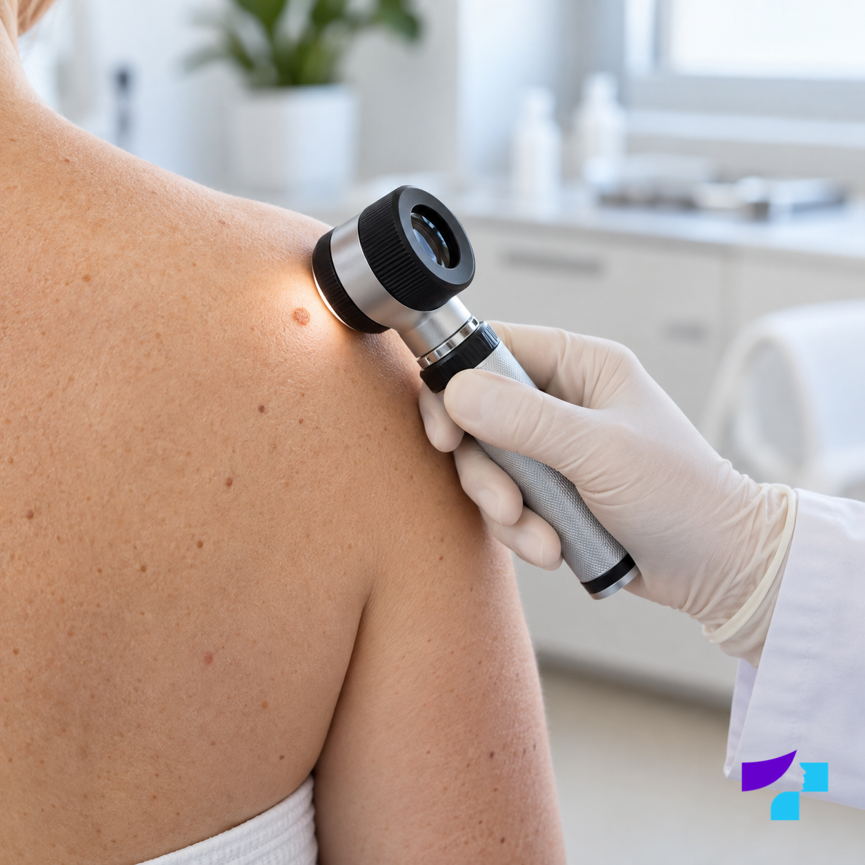
Examining a skin lesion with a dermatoscope allows the doctor to assess structures and pigmentation under magnification that cannot be fully seen with the naked eye.
The doctor assesses the lesion’s appearance, location, duration, changes over time and symptoms, as well as the patient’s age, skin type, sun exposure, family history and previous skin conditions. Dermoscopy, examination of the lesion under magnification and special lighting, is often used.
Dermoscopy helps assess pigmented and non-pigmented lesions, moles, seborrhoeic keratoses, warts, actinic keratoses and suspicious skin tumours. It does not always provide the final diagnosis: the doctor also considers the clinical examination and medical history, and suspicious cases require tissue examination.
Skin mapping and digital monitoring
A dermatologist may recommend digital documentation or skin mapping for some patients with many moles or an increased risk of melanoma. Lesions and their images are recorded so that later visits can establish whether a lesion has changed or a new one has appeared.
This approach may help people with many moles, atypical naevi, previous melanoma, a family history of melanoma or lesions in areas that are difficult to inspect, such as the back. A doctor may set a shorter follow-up interval for an individual unclear lesion. When convincing malignant features are present, digital monitoring must not delay biopsy or removal.
Digital monitoring does not replace medical assessment, but it can make changes over time easier to evaluate objectively. Read more about the methods and individual risk assessment in the articles on dermoscopy and mole checks.
When are biopsy and histology needed?
A biopsy is the removal of a tissue sample for examination. Histology is the microscopic examination of tissue, which helps confirm a diagnosis and determine whether a lesion is benign, precancerous or malignant. Both a biopsy sample and a completely removed lesion can be examined histologically.
Histology may be needed when a lesion looks suspicious, changes, bleeds, ulcerates, cannot be diagnosed confidently or is being removed for diagnostic reasons. If melanoma or another skin cancer is suspected, the lesion should not be destroyed solely for cosmetic reasons without appropriate assessment and the opportunity to examine the tissue.
Removal of skin lesions
How is the removal method chosen?
The removal method depends on the diagnosis, size, depth and location of the lesion, the cosmetic risk and whether tissue is needed for histology. Depending on the lesion, options may include:
- surgical excision;
- shave excision;
- laser therapy;
- cryotherapy, or treatment with cold;
- electrocoagulation;
- curettage, or scraping of the lesion;
- other methods suitable for the specific diagnosis.
The choice of method is not merely cosmetic. A suspicious lesion requires a diagnostically appropriate method that provides tissue for histological examination. If a lesion is clearly benign but causes cosmetic or functional problems, the doctor can recommend a suitable removal method. Not every benign lesion needs to be removed.
Read more about specific treatments in the articles on mole removal, wart removal and papilloma removal. A cyst or repeatedly inflamed subcutaneous lump requires separate assessment, as its treatment differs from removal of a superficial skin growth.
Healing and possible effects
Redness, a crust, tenderness, pigmentation changes or a scar may occur after removal. The risk of scarring depends on the method, the lesion’s location and size, individual healing and aftercare. The doctor’s instructions on wound care and sun protection should be followed after the procedure.
Can skin lesions be removed at home?
Unclear skin lesions should not be cut, burned, treated with caustic substances, frozen, squeezed or treated with unproven remedies at home. Doing so may cause scarring, infection, burns or pigmentation changes and may delay the diagnosis of a serious condition.
It is especially dangerous to attempt to remove a pigmented, bleeding, ulcerated, rapidly growing or changing lesion at home. An inflamed cyst should not be squeezed or cut either, as this can worsen inflammation and increase the risk of infection. A clear diagnosis is needed before removal.
Some confidently identified common warts may be treated at home with pharmacy products intended for warts, following the instructions and advice from a doctor or pharmacist. This is not appropriate for unclear lesions, moles or growths on the face or genitals. People with diabetes, impaired circulation or sensation, or weakened immunity should discuss the suitability of self-treatment with a doctor.
How often should skin lesions be checked?
The appropriate frequency is individual. A person without particular risk factors may only need self-monitoring and a dermatologist visit when a suspicious change appears. For people with many moles, atypical naevi, fair skin, frequent sunburn, a family history of skin cancer, previous melanoma or weakened immunity, the doctor may recommend regular checks, such as once a year or more often.
If a doctor has advised short-term monitoring, the recommended follow-up interval should be observed. A rapidly changing, bleeding, ulcerated or painful lesion should not be left until the next preventive visit.
How should you prepare for a dermatologist appointment?
Before the appointment, note when the lesion appeared and whether it changes, bleeds, itches, hurts, crusts or has been injured. Previous photographs may be useful. Decorative cosmetics should not be applied directly to the lesion before the visit.
Questions to ask the doctor include:
- What is this lesion most likely to be?
- Is dermoscopy needed?
- Are biopsy or histology needed?
- Can the lesion be monitored, or would removal be preferable?
- Which removal method is most suitable?
- Could the subcutaneous lump be a cyst, lipoma or another lesion?
- Could the thickened area be a scar, keloid or another lesion?
- Is digital monitoring needed for multiple lesions?
- What is the risk of scarring or pigmentation changes?
- How often should my skin be checked in future?
Common myths about skin lesions
| Myth | Fact |
|---|---|
| All skin lesions are dangerous | Many are benign, but changing or atypical lesions should be checked. |
| A benign lesion can never change | Benign lesions may become inflamed or injured, but changes still need assessment. |
| A wart can always be recognised by its appearance | Other lesions, including skin tumours, may resemble a wart. |
| All papillomas are caused by HPV | Skin tags, which are often called papillomas in everyday language, are not the same as HPV-related warts. |
| A cyst can simply be squeezed | Squeezing can promote inflammation and infection; the cyst may recur if its wall remains. |
| A mole can be removed with a laser without examination | A mole should be assessed by a dermatologist before removal. Destroying a suspicious lesion may prevent histological examination. |
| A red lesion is always an angioma | A red lesion may also be inflammation or a malignant skin tumour. |
| A scar is always only a cosmetic concern | Some scars hurt, itch or grow, and scar-like changes sometimes need to be distinguished from other skin conditions. |
| Bleeding always means cancer | No, but bleeding without a clear injury is a warning sign. |
| Itching always means an allergy | Itching may also be a symptom of inflammation, irritation or a tumour. |
| Urticaria is a persistent skin growth | Individual wheals are usually temporary even when the condition itself lasts longer. |
| Psoriasis is contagious | Psoriasis is not an infection and is not contagious. |
| Folk remedies are safe | They can damage the skin and delay diagnosis. |
| Removal always leaves a large scar | The risk and appearance of a scar vary and depend on the method, lesion depth and healing. |
| Dermoscopy is only needed for moles | It is also used to assess many other skin lesions. |
Summary
Skin lesions vary widely. Many are benign, but some may be precancerous changes or skin cancer. They may be visible on the surface or palpable beneath the skin, as with cysts and lipomas. Patients may also use the term for papules, spots, hives, psoriasis plaques, dermatitis rashes or scars, so a persistent lesion needs to be distinguished from a temporary, inflammatory or post-injury skin change.
The most important task for the patient is not to make an exact diagnosis, but to notice changes over time. A lesion that grows, bleeds, ulcerates, does not heal, changes or looks atypical requires examination by a dermatologist. Dermoscopy, appropriately selected digital monitoring and, when needed, histology help determine whether observation, treatment or removal is appropriate.
Medical disclaimer
This information is provided for informational purposes and does not replace a consultation with a dermatologist, diagnosis, or treatment. The significance of a skin lesion depends on its type, changes over time, the patient’s risk factors, and a medical assessment. Consult a dermatologist if a lesion grows rapidly, bleeds, ulcerates, does not heal, changes colour or shape, becomes painful, or appears unusual. Lesions of uncertain nature should not be cut, burned, cauterised, squeezed, or treated with unverified remedies at home.
References and authoritative sources
- DermNet: common benign skin lesions
- DermNet: dermoscopy and its use in assessing skin lesions
- DermNet: skin tags or acrochordons
- DermNet: keloids and hypertrophic scars
- American Academy of Dermatology (AAD): signs and changes in moles that deserve attention
- American Academy of Dermatology (AAD): diagnosis and treatment of warts
- American Academy of Dermatology (AAD): overview of actinic keratosis
- Skin Cancer Foundation: melanoma warning signs and images
- MedlinePlus, a resource of the U.S. National Library of Medicine: skin biopsy, procedure and results
