A mole check is an assessment of skin lesions performed by a dermatologist. Its purpose is to distinguish benign skin lesions from those that require further diagnostics, monitoring or removal with histological examination. In everyday language, patients most often use terms such as “mole”, “suspicious mole” or “dark spot”, but in dermatological practice, doctors assess not only classic pigmented naevi, but also other pigmented and non-pigmented skin lesions that may visually resemble moles.
Most moles are benign and do not pose a health risk during a person’s lifetime. However, some malignant skin tumours, especially melanoma, may initially look similar to an ordinary mole or may develop as a new, seemingly insignificant skin lesion. For this reason, a mole check is not merely a cosmetic procedure. It is a preventive and diagnostic medical consultation that helps detect changes that cannot always be reliably interpreted with the naked eye.
It is especially important to understand that a suspicious mole does not always mean melanoma, but suspicious signs should not be ignored. It is difficult for a patient to reliably distinguish a benign mole from a dysplastic naevus, seborrhoeic keratosis, pigmented basal cell carcinoma, precancerous changes of squamous cell carcinoma or early-stage melanoma. This is why mole diagnostics should be entrusted to a dermatologist.
What is a mole check?
A mole check is a structured assessment of skin lesions by a dermatologist or dermatovenerologist. During the consultation, the doctor listens to the patient’s concerns, assesses risk factors, examines the specific mole or several skin lesions and, if necessary, uses dermoscopy — examination of a skin lesion with a special magnifying device and light source.
The purpose of a mole check is not to “find cancer” in every mole. Its purpose is to determine whether a skin lesion appears benign or whether it has features that require monitoring, additional investigation, surgical removal or histological examination. Histology means microscopic examination of a removed tissue sample in a laboratory, which can provide a definitive diagnosis in suspicious cases.
What is mole diagnostics?
Mole diagnostics is a broader process than simple visual inspection. It combines several steps: taking the patient’s medical history and assessing risk factors, clinical examination of the skin lesion, dermoscopic assessment, comparison with other moles on the body and a decision on further management.
A dermatologist evaluates not only whether a mole is dark or large. Its shape, borders, colour uniformity, surface structure, growth, bleeding, itching, tenderness, ulceration and whether it differs from the patient’s other moles are all important. Sometimes the most suspicious lesion is not the largest or darkest one, but the one that looks different from the rest of the person’s moles.
Why do moles form and why can they change?
Moles, or melanocytic naevi, form from melanocytes — cells that produce melanin, the pigment that gives skin its colour. Moles may be congenital or acquired during life. Some appear in childhood and adolescence, while others become more noticeable due to hormonal changes, sun exposure or skin ageing.
Moles may become darker or lighter, slightly raised above the skin, or change as the body grows. Not every change means a malignant process. However, rapid, uneven or unexplained changes should be assessed carefully, especially in adulthood.
This is not an infectious disease. Moles are not contagious, they cannot be caught from another person, they do not spread from person to person, and they do not have an “incubation period”. Therefore, this article does not include an infection mechanism or infection timeline. In the context of mole checking, the key issue is another mechanism — how UV radiation, genetic predisposition, skin type, number of moles and immune status can influence skin cell damage and the risk of malignant changes.
How does the risk of malignant changes arise?
Skin cells are exposed to environmental influences every day. One of the most important risk factors for skin cancer is ultraviolet radiation — both from the sun and from artificial UV sources, such as tanning beds. UV radiation can damage cellular DNA, the genetic information that regulates cell growth and renewal. If this damage accumulates and the body cannot fully repair it, uncontrolled growth may begin in some cells.
Melanoma develops from melanocytes — pigment cells. It may appear in an existing mole, but it often develops on previously normal skin. This is important because patients should not monitor only moles they have known for a long time. Attention should also be paid to new pigmented or unusual lesions, especially if they change rapidly in adulthood.
The skin “remembers” sun exposure. Repeated sunburns, prolonged exposure to direct sunlight without protection and tanning bed use can increase the risk of skin cell damage. These changes do not always become visible immediately. Sometimes skin changes appear only after a longer period of time, which is why mole checks are important even when there is no pain or discomfort at the time.
Who is at higher risk?
A mole check is recommended for anyone who notices a new, changing or unusual skin lesion. However, there are groups of people for whom skin lesion monitoring is especially important.
Higher risk may apply to people who have many moles, atypical or dysplastic moles, fair skin, light-coloured eyes, fair or red hair, skin that burns easily in the sun, repeated sunburns during life, especially in childhood or youth, a history of tanning bed use, melanoma or another skin cancer in themselves or in first-degree relatives, weakened immunity or long-term immunosuppressive therapy. People who work outdoors, spend a lot of time in direct sunlight or have received a high lifetime dose of UV radiation should also be more cautious.
A risk factor does not mean that a person will definitely develop melanoma or another skin cancer. It means that skin lesion checks and sun protection become more important.
When should a mole be checked?
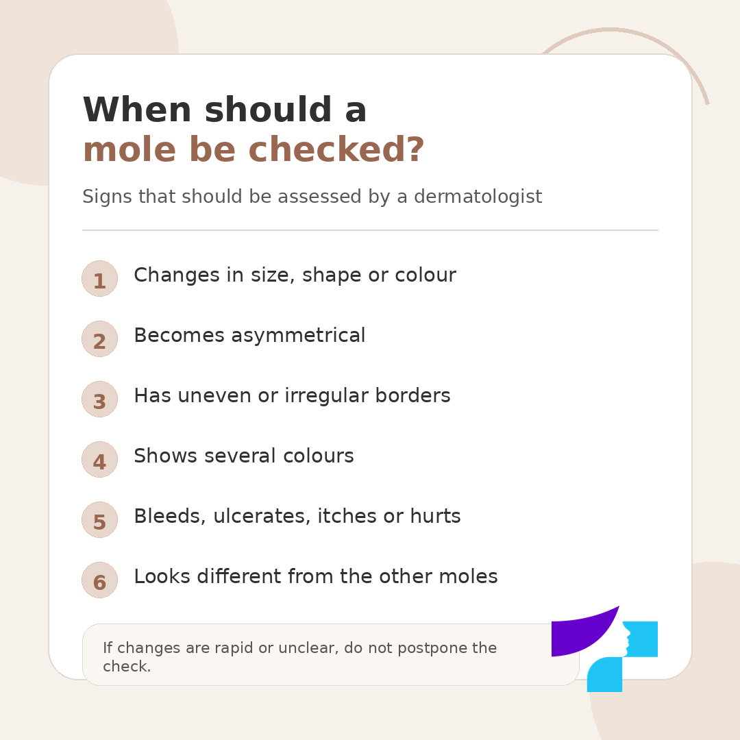
A mole should be checked if it changes in size, shape or colour, becomes asymmetrical, develops uneven borders, starts itching, hurting, bleeding, forming crusts, ulcerating or becoming raw. A dermatologist’s examination is also needed if a new unusual skin lesion appears, especially in adulthood, or if one mole clearly differs from the others.
It is especially important not to wait if changes occur rapidly — over weeks or a few months. Some malignant skin lesions do not cause pain or discomfort at first, so “no pain” does not mean that a check is unnecessary.
Three to five main signs that may indicate dangerous changes
Patients do not need to diagnose themselves at home, but they should know which signs are reasons to see a doctor. The most important signs are: a mole changes rapidly, becomes asymmetrical or develops irregular borders; several colours appear in it or the colour becomes uneven; the mole bleeds, ulcerates, itches or hurts without a clear reason; the lesion looks different from the other moles; a new atypical pigmented lesion appears in adulthood.
What does a suspicious mole look like?
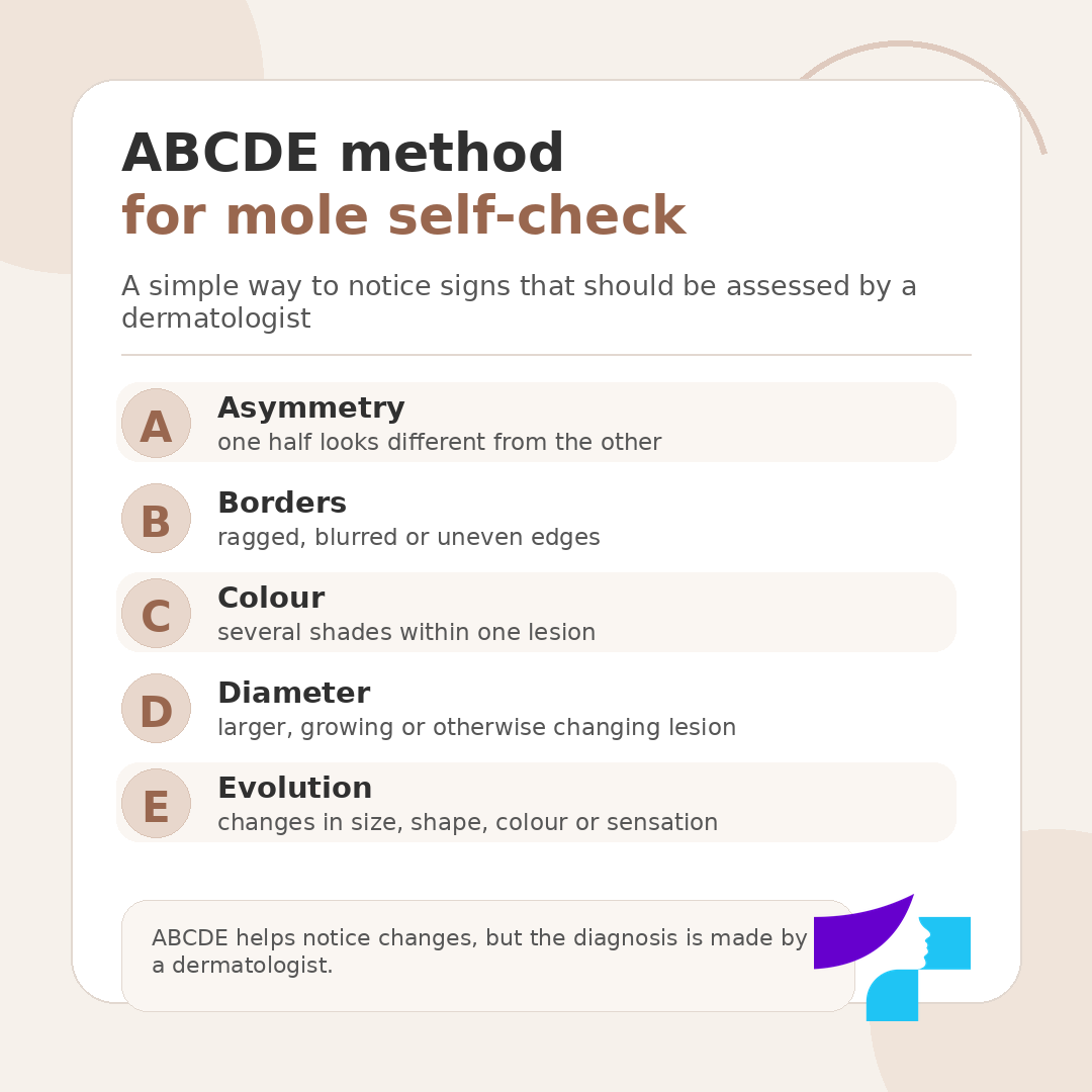
A suspicious mole is often explained using the ABCDE principle. This is a simple self-check tool that helps notice signs that should be shown to a dermatologist.
A stands for asymmetry — one half of the mole differs from the other. B stands for borders — they are irregular, ragged, blurred or uneven. C stands for colour — there are several shades in one lesion, such as light brown, dark brown, black, pink, greyish, white or bluish. D stands for diameter — attention should be paid to larger or growing lesions, although a small lesion can also be important if it changes. E stands for evolution — the mole changes over time in size, shape, colour, surface or causes new sensations.
The ABCDE method is not perfect and does not cover all possible forms of melanoma. Some melanomas may be light, pink, subtle or may not match the classic image of a dark mole. Therefore, self-checking helps notice changes, but it cannot replace dermatological diagnostics.
What is the “ugly duckling” sign?
The “ugly duckling” sign means that a lesion may be suspicious if it looks different from the other moles on that person’s skin. In some people, moles are light brown and similar to one another; in others, they are darker or raised. If one mole stands out — it is darker, lighter, pinker, larger, differently shaped or changing faster — it should be checked.
This principle is especially useful for people with many moles. The most suspicious lesion is not always the largest one. Sometimes the most important warning sign is that it differs from the person’s usual “mole pattern”.
How to check moles at home?
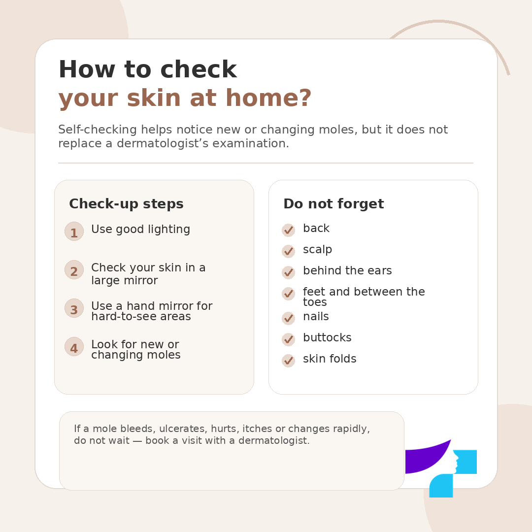
Skin self-examination helps detect a new or changing lesion in time, but it does not replace a dermatologist’s examination. It is best to perform a self-check in good lighting, using a large mirror and, if necessary, a hand mirror. A family member or another close person may also help, especially when checking the back, scalp and other hard-to-see areas.
First, examine the face, neck, chest, abdomen and arms. Then check the sides of the body with arms raised, the forearms, palms, spaces between the fingers and nails. Continue with the legs, tops and soles of the feet and spaces between the toes. It is especially important not to forget the back, buttocks, scalp, back of the neck and areas behind the ears.
During self-examination, pay attention to the ABCDE signs, the “ugly duckling” sign and any changes that were not present before. If a mole becomes different, bleeds, ulcerates, itches, hurts or grows rapidly, it should not simply be photographed and watched — a medical examination is needed.
Which body areas are most often forgotten during self-checks?
Many people check only the most visible areas — the face, arms, chest or abdomen. However, suspicious skin lesions can also appear in places that are rarely examined in daily life. Therefore, during self-checks and dermatological examinations, it is important not to forget the back, back of the shoulders, scalp, areas behind the ears, back of the neck, feet, soles, spaces between the toes, nails, buttocks and skin folds.
If a patient cannot check the back or scalp themselves, a close person may help; a hairdresser may also notice a lesion on the scalp, but the final assessment is always made by a doctor. Any new or changing lesion in a hard-to-see area is just as important as a mole on the face or arms.
Are all suspicious moles melanoma?
No. Many suspicious-looking moles turn out to be benign. Sometimes the cause of concern is seborrhoeic keratosis, an angioma, dermatofibroma, papilloma, wart, inflamed mole or mechanically traumatised skin lesion. However, these lesions can visually resemble more dangerous changes, so self-diagnosis may be misleading.
The dermatologist’s task is to assess whether the lesion is benign, whether it should be monitored, photographed for comparison, removed or sent for histological examination.
Does melanoma always look like a dark mole?
No. Classically, melanoma is often described as a dark, uneven, changing pigmented lesion, but it may also be pink, light-coloured, greyish, bluish, nodular, ulcerated or less noticeable. In people with darker skin, suspicious lesions may also appear on the palms, soles, under the nails or in areas that are not often examined in daily life.
Therefore, colour is not the only important sign. Changes over time, difference from other lesions and any new symptoms — bleeding, ulceration, itching, pain or rapid growth — must also be assessed.
How is a mole checked by a dermatologist?
A mole check usually begins with a conversation. The doctor asks when the lesion was noticed, whether it has changed, whether there has been bleeding, itching, pain, trauma, sunburn, tanning bed use, a family history of skin cancer or previous removal of skin lesions.
The dermatologist then examines the specific mole or several skin lesions. Depending on the purpose of the visit, the doctor may focus only on the lesion indicated by the patient or may recommend a broader skin examination. If the lesion appears suspicious or requires more precise assessment, a dermatoscope is used. Dermoscopy is mentioned in this article only as part of a mole check; a detailed explanation of optical, digital and video dermoscopy should be included in a separate article about dermoscopy.
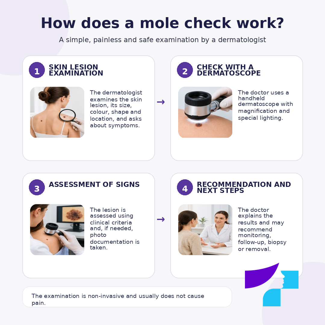
At the end of the visit, the doctor explains the conclusion. Possible outcomes include: the lesion appears benign and self-monitoring is sufficient; the lesion should be rechecked after a certain period of time; removal is recommended; histological examination is required; or the patient should be referred to another specialist if broader oncological or surgical management is needed.
What does the patient receive after a mole check?
After a mole check, the patient receives the doctor’s explanation of the nature of the examined lesion and further management. The doctor explains whether the lesion appears more likely to be benign, suitable for monitoring or suspicious, and indicates when a repeat check or additional examination is needed.
Depending on the situation, the doctor may recommend self-monitoring, a repeat visit after a certain period, lesion removal, histological examination or an additional consultation with another specialist. If dermoscopic assessment has been performed, the doctor explains why the specific lesion is considered calm or why it requires further monitoring.
Does the dermatologist check only one mole or the whole body?
This depends on the purpose of the visit and the patient’s risk factors. If the patient comes with one specific mole, the doctor may focus on that lesion. However, if the patient has many moles, has previously had skin cancer, has a family history of melanoma or has several suspicious lesions, the dermatologist may recommend a broader skin examination.
A full-body skin examination is especially useful if a person has many pigmented lesions and cannot regularly check the back, scalp, buttocks, back of the legs or other hard-to-see areas themselves.
How often should moles be checked?
There is no single check-up interval that suits everyone. For a person without increased risk, regular self-examination and a dermatology visit if a new or changing lesion appears are usually sufficient. For people at increased risk, the dermatologist may recommend periodic checks, for example once a year or at another individually determined interval.
For many people, skin self-examination approximately once a month is a practical guideline, especially if there are many moles or a history of intense sun exposure. However, the frequency of medical checks is determined individually, taking into account the number and type of moles, skin type, previous sunburns, family history, age, immune status and previous examination results.
If the dermatologist recommends a repeat check after a specific period, this interval should be followed, because some lesions are more safely assessed over time — by comparing whether they change.
Does a mole check hurt?
No. Mole examination and dermoscopic assessment are painless and non-invasive. Non-invasive means that the skin is not cut or damaged. The doctor may touch the skin with a dermatoscope or examine the lesion from a short distance, depending on the type of device used.
Pain may occur only if removal or biopsy is performed later, but local anaesthesia is usually used for such procedures. The mole check itself is not painful.
Should a mole be checked before removal?
Yes. Before removing a mole or another pigmented skin lesion, it should be assessed by a dermatologist. This is important for two reasons. First, the doctor determines whether the lesion appears benign or suspicious. Second, this determines the most appropriate removal method and whether histological examination is needed.
A suspicious pigmented lesion should not be removed only cosmetically, for example by destroying its superficial part without the possibility of fully examining the tissue. If there is suspicion of a malignant process, it is important to obtain adequate material for histology and choose the correct medical approach.
Monitor or remove a mole?
Not all moles need to be removed. Some benign lesions can simply be monitored, especially if they do not change, cause no symptoms and do not appear suspicious on dermoscopy. In such cases, the doctor may recommend self-examination, a repeat check after a certain period or photographing the lesion for comparison.
Removal may be recommended if the lesion looks suspicious, changes rapidly, bleeds, ulcerates, is repeatedly traumatised, bothers the patient or if histological confirmation of the diagnosis is needed. In suspicious pigmented lesions, it is important to choose a removal method that allows the tissue to be sent for histological examination.
Can a mole be removed with a laser?
The mole removal method should be chosen only after medical assessment. Laser treatment may be suitable for certain benign skin lesions, but suspicious pigmented lesions should not simply be “burned off” or cosmetically removed without diagnostics. The main risk is losing the possibility of full histological examination or delaying the detection of a malignant process.
If a mole is suspicious, the doctor usually chooses a method that allows the tissue to be sent for histology. The patient should not choose a removal technique based only on “faster” or “no scar”. Safety and diagnostic accuracy are more important than cosmetic convenience.
What happens if a mole turns out to be suspicious?
A suspicious finding does not yet mean that the patient has melanoma or skin cancer. It means that the doctor sees features that need closer assessment. Depending on the situation, the dermatologist may recommend a repeat check after a certain period, digital documentation for comparison, mole removal with histological examination or biopsy.
If the doctor considers that the mole has a high risk of malignant changes or if its nature cannot be reliably determined after examination and dermoscopic assessment, mole removal and/or biopsy may be recommended. Biopsy means taking a skin tissue sample and sending it for histological examination to determine in the laboratory whether the lesion is benign or malignant. In suspicious pigmented lesions, the doctor chooses a diagnostic and removal approach that provides sufficient tissue material for an accurate diagnosis.
If histology confirms a malignant lesion, the further treatment plan is determined according to the type, depth, location and stage of the lesion. In such cases, surgical treatment and involvement of a team of specialists may be required. Specific treatment is determined by a doctor; patients should not attempt to treat the lesion themselves with ointments, acids, folk remedies or mechanical removal at home.
Can mole checks help detect skin cancer early?
Yes. The key value of mole checks is the ability to notice suspicious changes in time. Skin cancer outcomes are generally better when a lesion is detected early, before it has grown deeply into tissues or spread further.
Patients should understand that early diagnosis does not mean only visiting a doctor once every few years. It begins with paying attention to one’s skin: regular self-checks, noticing changes and seeing a dermatologist in time.
Mole checks in children
Moles can be checked in children in the same way as in adults. In childhood, moles are often more dynamic than in adults — they may appear newly, grow as the child grows, change shade, become more noticeable or sometimes seem less pronounced. This does not always mean disease.
However, a child’s mole should be checked if it grows rapidly, becomes asymmetrical, starts bleeding, ulcerating, itching or hurting, develops several colours or differs significantly from other skin lesions. A dermatologist’s examination is also needed if the lesion is repeatedly traumatised, for example by clothing, a comb or during sports activities.
Parents should not try to remove a child’s mole themselves or treat it with irritating substances. If there is doubt, it is safer to show the child’s skin lesion to a doctor than to monitor it for a long time only by comparing photographs online.
Moles during pregnancy
During pregnancy, hormonal changes may make the skin more pigmented, and some skin lesions may become more noticeable. However, pregnancy should not be used as an explanation for every mole change. If a lesion changes rapidly, bleeds, ulcerates, becomes painful or looks suspicious, it should be checked by a dermatologist.
Pregnancy is not a reason to delay assessment of a suspicious lesion. The doctor will choose the safest diagnostic and further management plan for the situation.
Moles after sun exposure and tanning bed use
After intense sun exposure, the skin may become darker, irritated or sunburned. Moles may appear more noticeable, but special caution is needed if after sun exposure a lesion changes shape or colour, starts bleeding, hurting or ulcerating. Sunburn and long-term UV exposure are important risk factors for skin damage.
Tanning beds are not a safe alternative to the sun. Artificial UV radiation also damages the skin and increases the risk of skin cancer. People who have used tanning beds, especially regularly or at a young age, should monitor skin changes more carefully and discuss suitable check-up frequency with a dermatologist.
Mole check before and after summer
A mole check can be performed at any time of year, but before summer it is especially useful for people with many moles, fair skin, previous sunburns or other risk factors. A check before the active sun exposure season helps assess existing skin lesions and receive individual advice on sun protection.
After summer, a check is useful if there has been intense sun exposure, sunburn or new changes in the skin. However, a suspicious mole should not be left until the end of the season. If a lesion changes, bleeds, ulcerates, itches, hurts or grows rapidly, it should be checked immediately.
Can the body develop immunity against melanoma or dangerous moles?
The body does not develop immunity against moles or melanoma in the same way as it may after some infectious diseases or vaccination. Moles are not an infection, and the body does not “learn” to fully protect the skin from new moles or malignant changes.
The immune system is involved in recognising damaged and abnormal cells, and in modern oncology immunotherapy is one of the treatment approaches in certain melanoma situations. However, this is not the same as natural protection against malignant changes in moles. In prevention, the key role belongs to reducing UV exposure, skin self-examination, awareness of risk factors and timely dermatological assessment.
How to prepare for a mole check?
Before the visit, it is useful to think about which mole or skin lesion is causing concern and when the changes were noticed. If possible, earlier photographs showing the lesion can be prepared, as they help assess the dynamics of changes. If dermoscopy, mole removal or histology has been performed previously, it is advisable to bring the reports or results.
On the day of the visit, decorative cosmetics, self-tanning products or thick creams should not be applied to the area being examined, as they may interfere with skin assessment. If a broader skin examination is planned, comfortable clothing that is easy to remove is preferable. Dark nail polish should be removed if there is suspicion of a lesion under or around the nail.
Can photographing a mole help?
Photography can help notice changes, especially if the mole is located in an area that is difficult to examine daily. However, home photographs have limitations: different lighting, distance, phone camera and angle may create the impression that a mole has changed or has not changed, even when the real situation is different.
If the dermatologist considers it necessary, the lesion can be documented medically for comparison over time.
Mole check price: what affects the cost?
The price of a mole check may vary depending on the doctor’s qualification, type of consultation, number of lesions examined and the extent of additional diagnostics required.
Can several moles be checked during one visit?
Yes, several moles or skin lesions can be checked during one visit. This is especially useful for patients who have many moles or are not sure which lesion is the most important. However, the duration and cost of the visit may depend on the number of lesions examined and the need for more detailed assessment.
If the patient has very many moles or requires systematic documentation of skin lesions, the doctor may recommend a separate examination plan or a repeat visit.
How to distinguish a mole from a wart, papilloma or seborrhoeic keratosis?
Patients often cannot do this reliably themselves. Seborrhoeic keratosis may look like a dark lesion “stuck” to the skin. Papillomas are often soft skin growths. Warts may be rough and related to human papillomavirus. Angiomas may appear as red vascular lesions. However, different benign and malignant lesions may sometimes visually overlap.
If a skin lesion changes, bleeds, hurts, ulcerates or looks atypical, it should not be identified only by comparing it with images on the internet. A dermatologist’s examination is safer than self-diagnosis.
What is medicine currently researching and developing in mole diagnostics?
Mole diagnostics is developing rapidly, especially in the field of skin imaging. In practice, digital documentation of lesions, comparison over time and full-body skin mapping in high-risk patients are becoming increasingly important. This helps doctors detect new or changing moles, especially in people with a large number of lesions.
Artificial intelligence and machine learning methods for analysing images of skin lesions are also being actively studied. These technologies may help sort images, detect suspicious lesions and support the doctor’s decision, but they do not replace a dermatologist. AI systems depend on image quality, datasets and clinical context. Therefore, the safest approach remains clinical assessment by an experienced doctor, using technologies as support tools rather than as a substitute for self-diagnosis or medical evaluation.
What can be done for prevention?
Not all skin cancer risk situations can be prevented, because some are influenced by genetics, skin type and family history. However, there are several practical steps that reduce the risk of skin damage caused by UV radiation.
It is advisable to avoid sunburn, avoid tanning beds, use broad-spectrum sunscreen, wear a hat, sunglasses and protective clothing, stay in the shade during periods of stronger UV radiation and check the skin regularly. Children’s skin should be especially protected from sunburn, because early and intense UV exposure may affect skin risks later in life.
When should a doctor be consulted urgently about a mole?
A dermatologist’s visit should not be delayed if a mole or another skin lesion changes rapidly, bleeds without a clear injury, repeatedly forms crusts, becomes painful, starts ulcerating, grows quickly, becomes markedly asymmetrical or differs significantly from other skin lesions. An urgent check does not mean that the diagnosis will necessarily be serious. It means that the lesion must be professionally assessed.
Summary of mole checks
A mole check is one of the simplest ways to detect suspicious skin changes in time. It is a painless, quick and understandable visit for the patient, during which a dermatologist assesses the nature of skin lesions and determines whether monitoring, additional diagnostics or removal is needed. Self-examination, the ABCDE method and the “ugly duckling” sign help patients notice warning signs, but the diagnosis is made by a doctor.
If a mole changes, bleeds, itches, hurts, ulcerates, becomes asymmetrical or looks different from the others, it should not be postponed until “later”. Timely assessment helps avoid unnecessary anxiety in the case of benign lesions and allows earlier action if medically significant changes are found.
Disclaimer
The information in this article is intended for informational and educational purposes only and does not replace medical consultation, diagnosis or treatment. Self-diagnosis of moles and other skin lesions may be inaccurate, because benign and malignant lesions may look similar. If you notice a new, changing, bleeding, itchy, painful, ulcerating or otherwise suspicious skin lesion, consult a dermatologist listed below the article. In cases of severe or rapidly worsening symptoms, significant bleeding, a rapidly growing ulcerating lesion or other high-risk signs, medical help should be sought immediately.
References and authoritative sources
- American Academy of Dermatology Association: ABCDE signs for recognising melanoma
- NHS: symptoms of melanoma
- NHS: moles and suspicious changes
- Cancer Research UK: symptoms of melanoma
- National Cancer Institute: moles, dysplastic naevi and melanoma risk
- Centers for Disease Control and Prevention: reducing skin cancer risk
- Centre for Disease Prevention and Control of Latvia: UV radiation and skin cancer risk factors
- European Association of Dermato-Oncology: European guidelines for melanoma diagnosis and treatment
- PubMed: European interdisciplinary melanoma guidelines, diagnostics section
- DermNet NZ: skin self-examination, the “ugly duckling” sign and ABCDE signs
- DermNet NZ: ABCDEFG signs of melanoma
- The Skin Cancer Foundation: melanoma warning signs
- American Cancer Society: signs and symptoms of melanoma
