A mole, or melanocytic naevus, is usually a benign skin lesion formed by a cluster of pigment-producing cells called melanocytes. Moles may be present at birth or appear later in life, and they can vary in colour, size, shape, surface and elevation above the skin.
Most moles do not become malignant and do not need to be removed preventively. However, a new, changing, spontaneously bleeding, ulcerating or noticeably different skin lesion should be examined by a dermatologist. Self-checking can be supported by the ABCDE rule and the “ugly duckling” sign, but a diagnosis cannot be made reliably from appearance or a photograph alone.
The main practical principle is simple:
know your moles → check your skin regularly → notice a new or changing lesion → if there are warning signs, book a mole check with a dermatologist.
What is a mole?
In medical language, a mole is most often called a melanocytic naevus. Melanocytes are skin cells that produce melanin — the pigment that contributes to the colour of the skin, hair and eyes. When melanocytic cells cluster in one area of the skin, a naevus can form.
Moles may be:
- light or dark brown;
- black or bluish;
- pink or skin-coloured;
- flat or raised above the skin;
- smooth, rough or slightly warty;
- with or without hairs.
The word “naevus” is broader than the everyday word “mole”. There are also other types of naevi that are not formed by melanocytes. This article mainly discusses melanocytic naevi — the lesions people most often mean when they talk about pigmented moles.
Not everything called a mole is a melanocytic naevus
In everyday language, almost any brown, red or raised skin lesion may be called a mole. In reality, several different skin lesions can look similar.
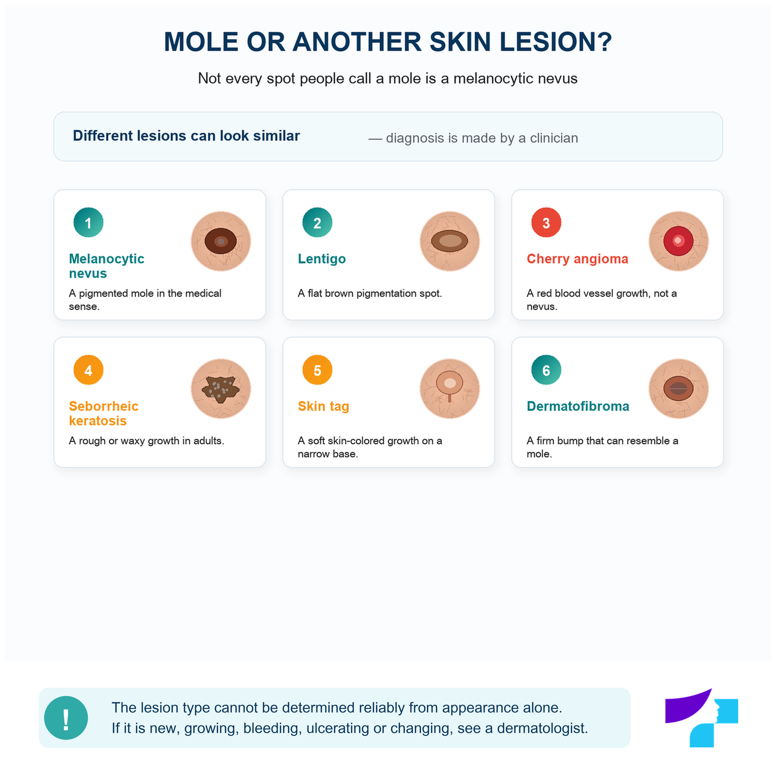
| Lesion | What can it look like? | Important note |
|---|---|---|
| Melanocytic naevus | Brown, black, bluish or skin-coloured; flat or raised | A mole in the medical sense |
| Lentigo | A flat, evenly brown patch | A pigment spot, not the same as a melanocytic naevus |
| Seborrhoeic keratosis | A brown, rough or waxy lesion that may look “stuck on” the skin | Common in adults and tends to increase with age |
| Angioma | A bright red or violet-red dot or small bump | A benign vascular lesion, not a melanocytic mole |
| Papilloma or skin tag | A soft skin-coloured lesion on a narrow base or “stalk” | Often appears in skin folds |
| Dermatofibroma | A firm brownish or pinkish nodule | May resemble a mole but is formed by other cells |
| Basal cell carcinoma or another skin tumour | A non-healing, shiny, pigmented, crusted or bleeding lesion | Requires a dermatologist’s assessment |
The type of lesion cannot be determined reliably from this table or from an online image. If a new, growing, bleeding or unclear skin lesion appears, professional assessment is needed.
Why do moles appear?
The number and appearance of moles are influenced by several factors.
Genetic tendency
People whose family members have many melanocytic naevi may also have more moles themselves. Genetic factors can influence the number, type and distribution of moles.
Age
Acquired moles most often appear in childhood, adolescence and early adulthood. The formation of new naevi usually decreases with age, but later in life other brown lesions, such as lentigines or seborrhoeic keratoses, may appear.
Ultraviolet radiation
Ultraviolet radiation from the sun can contribute to the formation of acquired moles and pigment changes, especially in childhood and adolescence. This does not mean that every mole is caused only by the sun.
Hormonal changes
During puberty and pregnancy, some moles may become slightly darker or more noticeable. Suspicious or rapid changes should not automatically be explained only by hormones.
Individual skin characteristics
The number, colour and visibility of moles may vary depending on skin type, pigmentation and individual characteristics.
Congenital and acquired moles
Congenital moles
A congenital melanocytic naevus is visible at birth or appears during the first weeks of life. It may be very small or cover a large area of skin.
As the child grows, a congenital mole usually enlarges proportionally with the body. This is not necessarily worrying. Medical assessment is needed if the lesion:
- grows much faster than the child;
- becomes unevenly nodular;
- changes colour significantly;
- bleeds or ulcerates;
- causes pain;
- becomes difficult to examine.
Large and giant congenital naevi require more careful monitoring. The follow-up plan depends on the expected adult size, location, appearance and other individual factors.
Acquired moles
Most moles are not visible at birth but appear later. New moles in children and teenagers are often a normal part of skin development.
A new pigmented lesion in adulthood is not automatically melanoma. However, it should be assessed more carefully, especially if it continues to grow, changes colour or shape, or clearly differs from the other lesions.
What are the common types of moles?
Common acquired melanocytic naevus
A common naevus is often round or oval, fairly symmetrical and evenly pigmented. It may be flat, slightly raised or clearly elevated above the skin.
Flat mole
A flat naevus usually looks like a brown patch that does not rise above the skin. A flat surface by itself does not prove that a lesion is benign.
Raised mole
Many benign naevi become raised over time. They may be soft, dome-shaped, light brown or skin-coloured. Elevation by itself is not a warning sign.
Atypical or dysplastic naevus
An atypical naevus may be larger, more asymmetrical, less sharply bordered or more unevenly pigmented than a common mole.
An atypical naevus is not the same as melanoma. Most atypical naevi are benign, but having several atypical moles can be associated with a higher melanoma risk. Not every atypical naevus needs to be removed automatically.
Blue naevus
A blue naevus is usually a small, smooth, bluish or blue-grey lesion. The colour appears because pigment is located deeper in the skin.
A typical blue naevus is usually benign and stable. A new, growing or changing blue-black lesion should be checked by a dermatologist.
Halo naevus
A halo naevus is a mole surrounded by an even white or noticeably lighter area of skin. This immune reaction is more common in children and young people, and the central naevus may gradually become lighter or disappear.
In an adult, a new white area around an unusual or changing mole should be assessed by a dermatologist.
Spitz naevus
A Spitz naevus is a benign melanocytic lesion that most often appears in children and young people. It may be pink, red, brown or dark, raised above the skin and may initially grow quite quickly.
A Spitz naevus can resemble melanoma clinically and, in some cases, microscopically. A rapidly growing or unusual nodule in a child should therefore be shown to a dermatologist.
What does a common mole look like?
A benign mole often has several of the following features:
- a round or oval shape;
- a fairly symmetrical appearance;
- clear and even borders;
- an even colour or several closely related shades;
- a size and shape that remain stable over time;
- no spontaneous bleeding, ulceration or non-healing crust.
There is no single universal appearance of a “normal” mole. Benign naevi may be flat or raised, light or dark, with or without hairs.
One isolated feature alone cannot confirm or rule out a malignant process.
How can moles change during life?
Moles are not completely unchanging lesions.
In childhood and adolescence, they may:
- appear for the first time;
- become larger as the child grows;
- change colour slightly;
- become raised above the skin.
In adulthood, some moles may become lighter or gradually disappear. After sun exposure they may look darker, while mechanical irritation can temporarily cause redness, tenderness or a crust.
Attention should be paid to a new, progressive or uneven change, rather than to the fact that a mole has changed slightly over many years.
What does a suspicious mole look like?
A new or existing skin lesion may be suspicious if it:
- has become asymmetrical;
- has developed irregular or blurred borders;
- has changed colour;
- contains several unevenly distributed colours;
- grows rapidly or unevenly;
- changes its surface or becomes nodular;
- bleeds spontaneously;
- ulcerates or repeatedly forms a crust;
- itches, is tender or painful for a prolonged time;
- looks clearly different from the person’s other moles.
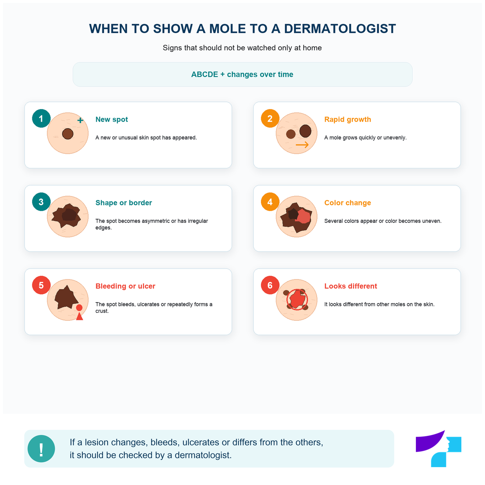
These signs do not mean that the lesion is definitely melanoma. They mean that it should be examined by a dermatologist. The most important melanoma warning signs are a new lesion, change in an existing lesion and a lesion that differs from the other skin spots.
The ABCDE rule for moles
The ABCDE rule helps assess a pigmented lesion in a structured way. It is a self-checking tool, not a diagnostic method.
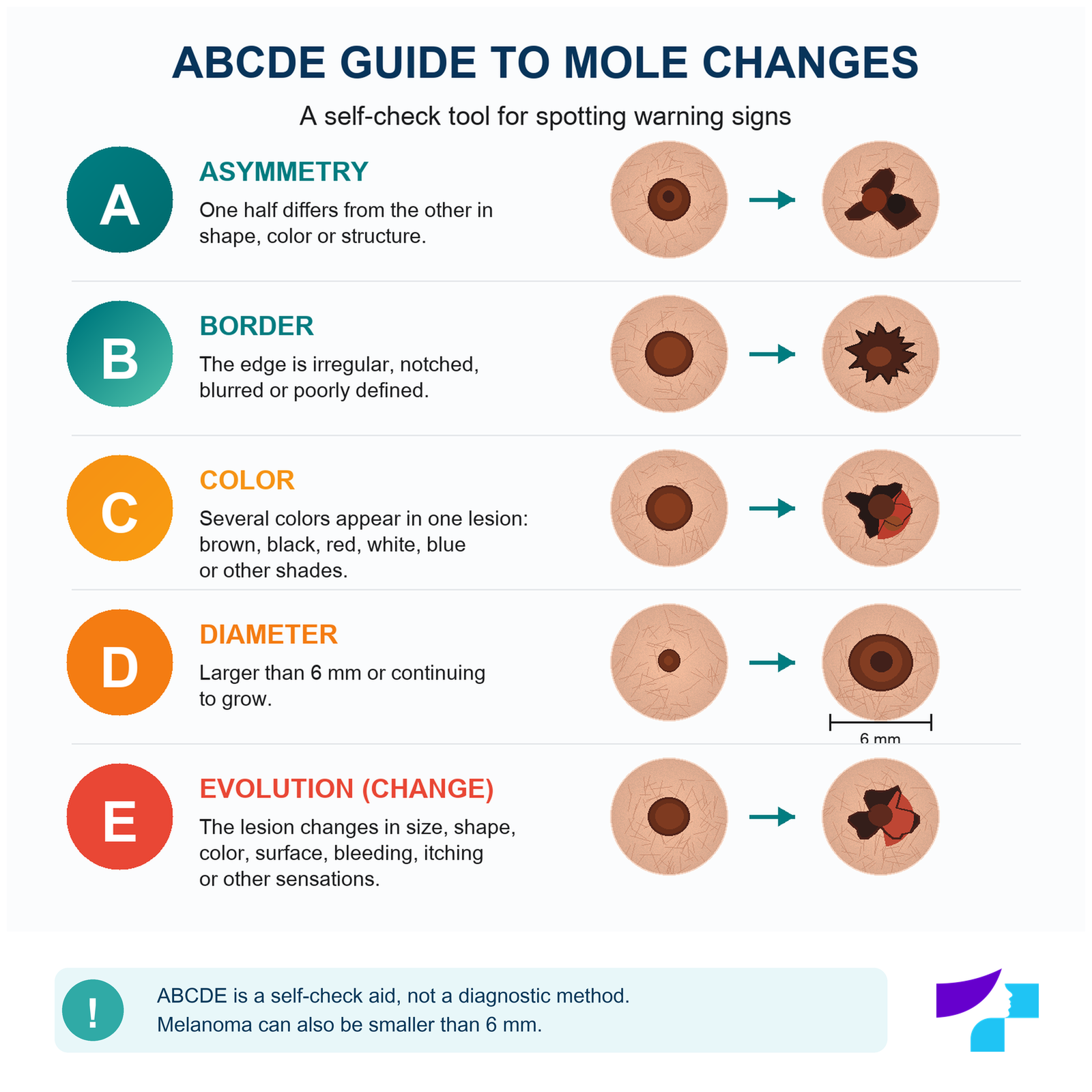
| Letter | Meaning | What can be noticed? | Important limitation |
|---|---|---|---|
| A | Asymmetry | One half of the lesion differs from the other in shape or colour | A benign naevus may also be slightly asymmetrical |
| B | Borders | The borders are jagged, blurred or poorly defined | Borders must be assessed together with other signs |
| C | Colour | Uneven brown, black, red, white or bluish shades appear within one lesion | Some benign naevi can have more than one shade |
| D | Diameter | The lesion is larger than about 6 mm or continues to enlarge | Melanoma can also be smaller than 6 mm |
| E | Evolution | Size, shape, colour, surface or sensations change over time | Change is often more important than one isolated sign |
Not every mole larger than 6 mm is dangerous, and not every melanoma is larger than 6 mm. Diameter is only one assessment criterion.
What is the “ugly duckling” sign?
A person’s moles often have a similar overall pattern. They may be similar in colour, size and shape.
The “ugly duckling” sign refers to a lesion that clearly differs from the others, for example:
- it is much darker or lighter;
- it is noticeably larger;
- it has a different shape;
- it is the only pink or nodular lesion;
- it changes while the other moles remain stable.
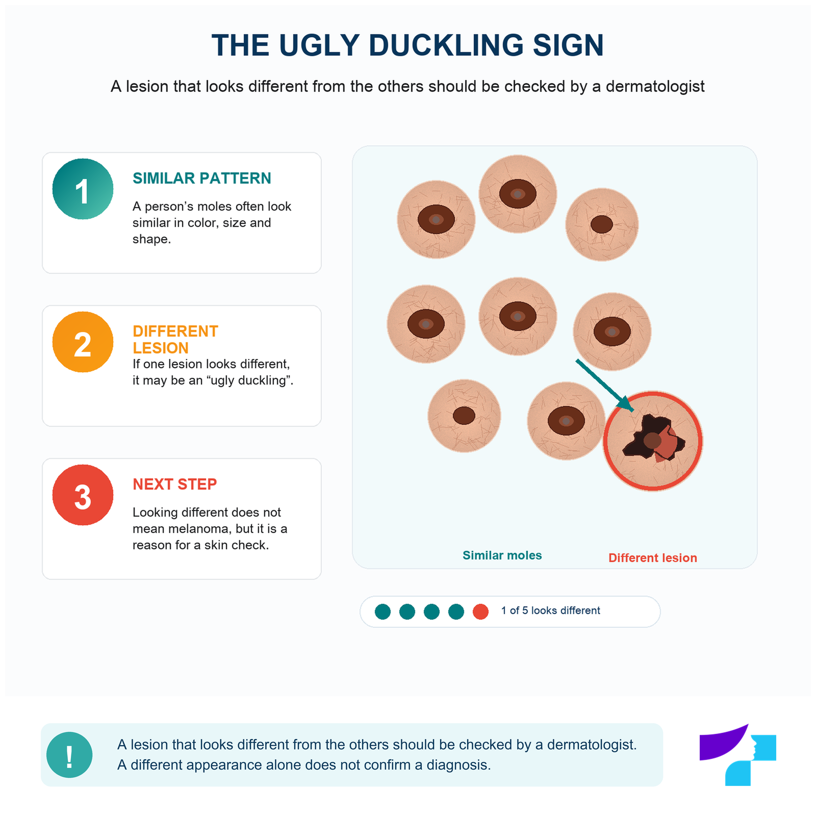
A different appearance does not automatically mean melanoma, but it is a valid reason for dermatological assessment.
Mole or melanoma?
Melanoma is a malignant tumour that develops from melanocytes. It may arise in a pre-existing mole, but many melanomas begin as a new lesion on previously normal-looking skin.
This means that existing moles are not the only lesions to watch. Any new or unusual patch or nodule should also be noticed.
Most moles do not become melanoma, so preventive removal of all naevi is not necessary. Knowing your skin, protecting it from the sun and checking suspicious lesions in time are far more important.
What does the colour and shape of a mole mean?
Dark or black mole
Dark colour by itself does not mean melanoma. A benign naevus may be dark brown or almost black.
Attention should be paid to whether the mole:
- has become significantly darker;
- has developed uneven colour;
- is changing;
- contains new black, bluish, red or white areas.
Bluish mole
A bluish colour can be caused by pigment located deeper in the skin, as in a blue naevus. A new or changing blue-black lesion should not be diagnosed based on colour alone.
Multicoloured mole
Several unevenly distributed colours can be a warning sign. However, some atypical but benign naevi may also be unevenly pigmented.
Red “mole”
A bright red or violet-red lesion is often an angioma — a benign vascular lesion rather than a melanocytic naevus.
A red lesion should be checked if it:
- grows quickly;
- bleeds spontaneously;
- does not heal;
- changes shape;
- is unclear in nature.
Several angiomas should not automatically be interpreted as a sign of liver, kidney, pancreatic or other internal organ disease.
Mole on a “stalk”
A lesion on a narrow base may be a raised intradermal naevus, but it is often a fibroma or papilloma. The exact diagnosis is made by a dermatologist.
Moles in special and hard-to-see locations
During a skin self-check, it is important not to look only at the face, hands and front of the body.
Scalp
Lesions on the scalp can be difficult to notice. The hair can be checked in sections using a mirror or help from another person.
Palms and soles
Melanocytic naevi and other pigmented lesions may also occur on the palms and soles. On the sole, melanoma may sometimes look like a new dark, pink or rapidly growing lesion.
Under the nails
A dark line under a nail may often be related to trauma or another benign reason, but it should be checked if the line:
- has appeared recently;
- is becoming wider;
- has uneven colour;
- extends onto the skin around the nail;
- is associated with nail splitting, lifting or a nodule.
Melanoma can also develop under a nail, so changing pigmentation should not be treated for a long time only as nail trauma or fungal infection.
Back and other hard-to-check areas
The back, buttocks, backs of the thighs and other hard-to-see areas can be checked with a hand mirror or help from another person.
What does it mean if a mole itches, hurts or bleeds?
Itchy mole
Short-term itching can be caused by dry skin, friction, irritation or inflammation. Itching alone does not confirm melanoma.
Persistent or repeated itching should be assessed, especially if the mole also changes in size, shape, colour or surface.
Painful mole
Pain may occur after trauma, shaving, friction or inflammation. If a mole hurts without a clear reason, the pain does not go away or the lesion is changing at the same time, dermatological assessment is needed.
Bleeding mole
If a mole bleeds after a clear injury, bleeding should be stopped by pressing clean gauze or a dressing on the area.
A dermatologist’s consultation is needed if:
- bleeding started without a clear injury;
- bleeding recurs;
- a non-healing crust forms;
- the mole ulcerates;
- the lesion changes appearance at the same time.
Crust or ulcer
A one-time crust may form after mechanical injury. Repeated crusting, oozing, ulceration or a wound that does not heal should not be considered a normal mole feature.
What to do if a mole is injured or torn?
A mole can be injured while shaving, by a fingernail, jewellery, clothing or during sport.
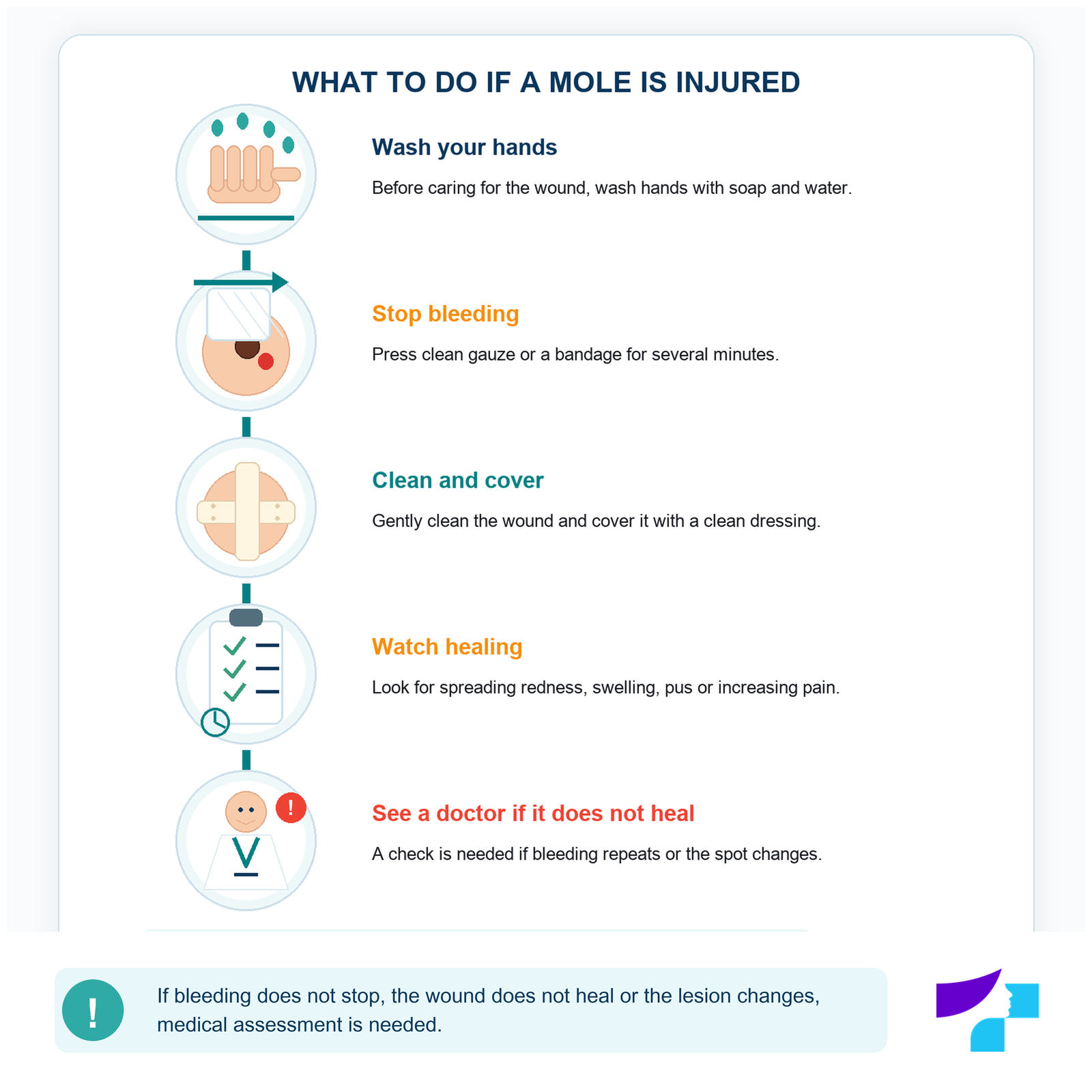
- Wash your hands.
- Apply clean gauze or a dressing.
- Use steady pressure to stop the bleeding.
- Then gently clean the wound.
- Protect it with a clean, non-stick dressing.
- Watch for increasing redness, swelling, pus or pain.
Do not try to cut off a partially torn mole, tie it with thread, burn it or treat it with wart-removal products.
A single injury by itself does not mean that a benign mole will become malignant. Trauma may cause bleeding and inflammation or draw attention to a lesion that had already been changing. If the wound does not heal, bleeding recurs or the mole continues to change, it should be checked.
Mole with hairs
A hair growing from a mole usually means that a hair follicle remains in that area. Hairs are common in benign naevi, but they are not an absolute safety sign.
Hair can be:
- carefully trimmed with clean scissors;
- shaved carefully without injuring the lesion.
Plucking a hair may irritate the hair follicle and cause redness or inflammation, so trimming is usually safer.
If a mole is repeatedly injured while shaving or rubs against clothing, a dermatologist can assess whether removal for practical reasons is justified.
Moles in children
New moles in childhood and adolescence are usually common. As a child grows, moles may gradually become larger, darker or lighter.
Parents do not need to check the child’s skin every day or worry about every new small mole. It is more important to notice a lesion that:
- grows much faster than the others;
- becomes noticeably larger than the child’s other moles;
- bleeds or ulcerates;
- becomes nodular;
- changes colour significantly;
- differs from the other lesions.
Melanoma in children is rare, but it does not always fit the classic adult ABCDE criteria. It may be pink or skin-coloured, fairly evenly coloured, raised above the skin or appear as a new rapidly growing nodule. In children, the evolution of a lesion and whether it differs from the others are especially important.
Protection from sunburn is particularly important in childhood.
Moles during pregnancy
During pregnancy, some women notice a change in mole colour or size. On the abdomen and breasts, a lesion may also look larger because the skin stretches.
Marked or uneven changes should not automatically be attributed to pregnancy. A dermatologist should examine a mole that:
- grows rapidly;
- becomes asymmetrical;
- changes colour;
- bleeds;
- ulcerates;
- clearly differs from other lesions.
Dermoscopy is a non-invasive examination method, so a suspicious mole can also be checked during pregnancy. Assessment should not be postponed until after childbirth.
Moles, sun and ultraviolet radiation
Ultraviolet radiation from the sun and tanning beds damages skin cells and increases the risk of skin cancer. Preventing sunburn in childhood and adolescence is especially important.
Sun protection should combine several measures:
- stay in the shade when ultraviolet radiation is intense;
- wear clothing that covers the body;
- wear a hat;
- apply a broad-spectrum sunscreen to exposed skin;
- use enough sunscreen and reapply it regularly;
- avoid tanning beds.
It is not necessary to cover every mole with a plaster. It is more important to protect the whole skin, not only individual pigmented lesions.
Sunscreen does not provide complete protection and is not intended to allow unlimited time in the sun.
How to check moles at home
Skin self-examination helps notice a new or changing lesion, but it does not replace a dermatologist’s assessment.
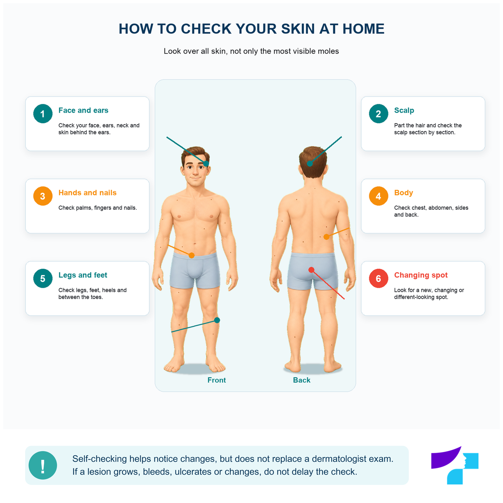
- Choose a well-lit room.
- Use a full-length mirror and a hand mirror.
- Check the face, ears, neck and scalp.
- Check the arms, palms, spaces between fingers and nails.
- Check the chest, abdomen and sides of the body.
- Use a mirror or help from another person to check the back and shoulders.
- Check the buttocks and the front and back of the legs.
- Check the feet, heels, spaces between toes and nails.
- Look for a new, changing or different-looking lesion.
There is no single self-check interval suitable for everyone. It should be adapted to individual risk factors and the dermatologist’s recommendations. If a suspicious change is noticed, do not wait for the next planned check.
How to photograph a mole for monitoring
A photograph can help compare a lesion over time.
- Take photographs in similar lighting.
- Make sure the image is in focus.
- Take both a close-up and a wider image of the body area.
- Save the date of the photograph.
- If needed, place a size reference nearby without touching the lesion.
- Do not use filters or colour correction.
A normal phone photograph does not show the structures that a dermatologist sees during dermoscopy. If the lesion is suspicious, photography should not replace medical consultation.
Who needs more careful mole monitoring?
Greater attention is needed for people who have one or more melanoma risk factors:
- very many moles;
- several atypical naevi;
- previously diagnosed melanoma;
- melanoma in a first-degree relative;
- fair skin, freckles and a tendency to sunburn;
- repeated sunburns;
- tanning bed use;
- weakened immunity or immunosuppressive treatment;
- a large or giant congenital melanocytic naevus;
- lesions in hard-to-see locations;
- visual or movement limitations that make full skin checks difficult.
Having risk factors does not mean that a person will definitely develop melanoma. It means that sun protection and individually planned dermatologist checks are more important.
How does a dermatologist check a mole?
The dermatologist asks:
- when the lesion was noticed;
- how it has changed;
- whether there has been bleeding, itching, pain or trauma;
- whether other moles have been removed before;
- whether the patient or family members have had melanoma;
- what sun and tanning bed exposure has been like.
The doctor may examine not only the mole indicated by the patient, but also the rest of the skin. Comparing lesions helps identify one that differs from the person’s usual mole pattern.
For a broader assessment of the skin and individual risk, see the Medart article on mole checks.
What is dermoscopy?
Dermoscopy is a non-invasive examination of a skin lesion under magnification and special light. It allows the dermatologist to see pigment, blood vessel and other structures that are not sufficiently visible to the naked eye.
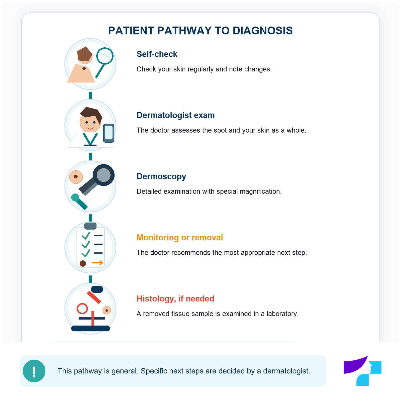
Dermoscopy helps decide whether a lesion:
- looks benign;
- should be monitored;
- should be photographed and compared over time;
- should be removed and sent for histological examination.
Dermoscopy is not a self-diagnosis method and does not always provide the final diagnosis. In certain cases, the diagnosis is confirmed by histological examination after the lesion is removed.
When is mole removal recommended?
Moles do not need to be removed simply because they exist. Removal may be considered if:
- there is suspicion of a malignant process;
- a precise diagnosis is needed;
- the lesion is repeatedly traumatised;
- it causes chronic irritation;
- it causes functional problems;
- there is a justified cosmetic reason after medical assessment.
A suspicious pigmented lesion should not be destroyed cosmetically without a dermatologist’s assessment. The doctor must choose a method that is medically safe and, if needed, allows histological examination.
For indications, methods and aftercare, see the article on mole removal.
Why should a mole not be removed at home?
Cutting, tying, burning or treating a mole with acids or wart-removal products can cause:
- bleeding;
- infection;
- a pronounced scar;
- incomplete removal;
- recurrent pigmentation;
- delayed diagnosis;
- loss of the opportunity for proper histological examination.
If a lesion is bothersome or repeatedly injured, the safest first step is to have it checked by a dermatologist.
Common myths about moles
“All moles larger than 6 mm are dangerous.”
Incorrect. Diameter is only one ABCDE feature. A large naevus may be benign, and melanoma may be smaller than 6 mm.
“A hair means the mole is completely safe.”
Incorrect. Hairs often grow in benign moles, but they are not an absolute safety sign.
“If a mole is torn, cancer develops.”
There is no reason to assume that a single injury automatically turns a benign naevus into melanoma. However, an injured lesion should be monitored and shown to a doctor if it does not heal or continues to change.
“Melanoma is always black.”
Incorrect. Melanoma may be brown, black, pink, red, bluish or almost skin-coloured.
“Melanoma always starts in a mole.”
Incorrect. Melanoma may develop in an existing naevus, but it often appears as a new skin lesion.
“All moles should be removed preventively.”
Incorrect. Most moles are benign. Preventive removal of all naevi is not necessary and does not replace skin self-checking.
“If a mole does not hurt, it is definitely safe.”
Incorrect. Early melanoma often causes no pain. Changes in the lesion may be more important than sensations.
When should you see a dermatologist?
A dermatologist consultation is needed if:
- a new and unusual lesion appears;
- a mole changes in size, shape or colour;
- it becomes asymmetrical;
- it develops irregular borders;
- it clearly differs from the other lesions;
- it bleeds spontaneously;
- it ulcerates or repeatedly forms a crust;
- it grows rapidly;
- it itches or hurts for a prolonged time;
- it is repeatedly traumatised;
- new or changing pigmentation appears under a nail;
- the person has several melanoma risk factors;
- the lesion causes justified concern.
When should the check not be delayed?
A dermatologist’s assessment should not be delayed if:
- the lesion has changed significantly within weeks or months;
- it bleeds without clear trauma;
- an ulcer or crust forms repeatedly;
- a wound does not heal;
- a new dark or multicoloured lesion appears;
- the mole differs clearly from the other lesions;
- pigment reappears in the scar of a previously removed mole;
- a dark streak under a nail changes or widens.
Mole changes are usually not a reason to call emergency medical services. More urgent help is needed if bleeding after trauma cannot be stopped, the wound is deep, or rapidly increasing pain, swelling, pus, spreading redness or fever develops.
How to prepare for a dermatologist consultation
Before the visit, it is useful to:
- write down when the change was first noticed;
- prepare earlier photographs if available;
- mention whether the lesion has bled, itched or hurt;
- tell the doctor about previously removed moles;
- bring previous histology results if available;
- mention melanoma or other skin cancer in the family;
- mention tanning bed use and sunburn history;
- avoid applying make-up directly on the lesion before the examination.
Medical disclaimer
The information in this article is intended for informational and educational purposes only and does not replace a dermatologist consultation, skin examination, dermoscopy, diagnosis or treatment. Moles, melanoma and other skin lesions may look similar, so a diagnosis cannot be made reliably from description, self-assessment or a photograph alone. If a lesion is new, changing, bleeding, ulcerating, painful, itchy or clearly different from the others, it should be examined by a dermatologist.
References and authoritative sources
- British Association of Dermatologists — patient information about melanocytic naevi, their causes, types and when assessment is needed
- DermNet — types, appearance, development and diagnosis of melanocytic naevi
- DermNet — congenital melanocytic naevi, growth in childhood and monitoring
- DermNet — melanocytic naevi and melanoma assessment during pregnancy
- American Academy of Dermatology — ABCDE warning signs of melanoma
- American Academy of Dermatology — skin self-examination guidance
- American Academy of Dermatology — possible signs of nail melanoma
- World Health Organization — ultraviolet radiation and skin cancer risk
Content author:
