A cafe-au-lait spot is a flat, light-to-medium brown patch of skin pigmentation. The name comes from French and means “coffee with milk”, because the colour may resemble milky coffee. Medically, this type of skin mark is often called a cafe-au-lait macule or CALM.
One or a few cafe-au-lait spots are often a benign skin feature. They are not an infection, are not contagious, are not a sign of poor hygiene and, by themselves, are usually not skin cancer. However, multiple cafe-au-lait spots, especially in a child, may be an important reason for medical evaluation, as they can sometimes be associated with genetic conditions such as neurofibromatosis type 1, Legius syndrome or McCune-Albright syndrome.
The key question is not only whether the spot is brown. A doctor evaluates the number, size, shape, borders, location, age of onset, family history and any associated signs.
Quick answers
| Question | Short answer |
|---|---|
| What is a cafe-au-lait spot? | A flat, evenly light-to-dark brown hyperpigmented patch on the skin. |
| Is it a mole? | Not necessarily. A cafe-au-lait spot is not the same as a melanocytic nevus, commonly called a mole. |
| Is it skin cancer? | Usually no. A typical isolated cafe-au-lait spot is not skin cancer and usually does not turn into it. |
| Is one cafe-au-lait spot normal in a child? | Often yes, if there are no other concerning signs. |
| When should I see a doctor? | If there are multiple spots, if the number increases, or if there are eye, nervous system, bone, developmental or hormonal signs. |
| Can the spot be removed? | Medical treatment is usually not needed. Laser treatment can be discussed for cosmetic reasons, but results are not guaranteed. |
What is a cafe-au-lait spot?
A cafe-au-lait spot is a localized area of skin hyperpigmentation. This means that a specific area of skin contains more melanin pigment than the surrounding skin. Melanin is the pigment that determines the colour of the skin, hair and eyes, and it is produced by skin cells called melanocytes.
A typical cafe-au-lait spot:
- is flat, not raised;
- is light brown, medium brown or dark brown depending on the person’s skin tone;
- usually has an even colour;
- may have clear, smooth or slightly irregular borders;
- does not cause itching, pain, burning or scaling;
- may be present at birth or appear in early childhood;
- often enlarges proportionally as a child grows.
The word “macule” in medicine means a flat skin spot that can be seen but usually cannot be felt as a raised lesion.
Why is it called a “coffee with milk” spot?
The term “cafe-au-lait” describes the colour of the spot, not its cause. On lighter skin, it often appears light brown or similar to milky coffee. On darker skin, it may be dark brown or only slightly darker than the surrounding skin.
The main feature is therefore not one exact colour, but a flat, evenly pigmented area that differs from the rest of the skin.
What does a cafe-au-lait spot look like?
A cafe-au-lait spot is most often an oval, round or irregular brown patch. It may be only a few millimetres in size or several centimetres across. Large spots may cover a wider area of skin.
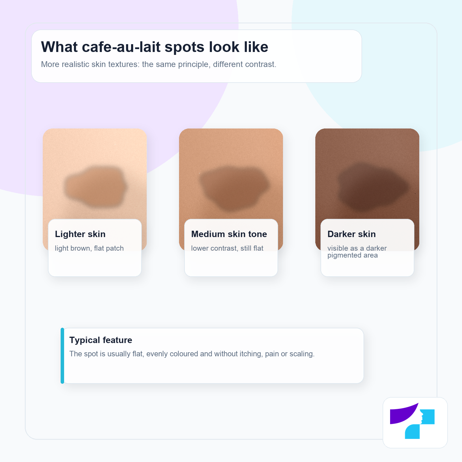
In dermatology, the descriptions “California coast” and “Maine coast” are sometimes used:
| Description | What it means | Clinical significance |
|---|---|---|
| Smooth, well-defined borders | The spot has a clearly demarcated, even edge | More often seen in isolated cafe-au-lait spots and in the context of NF1 |
| Irregular, jagged borders | The edge of the spot is uneven | May be seen in segmental pigmentation or McCune-Albright syndrome |
This comparison is not a diagnosis by itself. It is only one part of the clinical assessment.
Are cafe-au-lait spots dangerous?
One or a few cafe-au-lait spots are usually not dangerous. They are benign pigmented patches and most often do not cause health problems.
More attention is needed when there are many spots. Multiple cafe-au-lait spots, especially in a child, may be associated with a genetic syndrome. This does not mean that the child definitely has a serious condition, but it does mean that medical evaluation is appropriate.
Cafe-au-lait spots in children and infants
A cafe-au-lait spot may be visible at birth. Sometimes it is difficult to notice in a newborn and becomes more visible during the first years of life.
Parents may find it useful to note:
- how many spots there are;
- where they are located;
- their approximate size;
- whether new spots appear;
- whether there are other signs, such as freckle-like spots in the armpits or groin, lumps under the skin, vision problems, developmental delay or learning difficulties.
If a child has one small spot and no other symptoms, it is often simply observed. If there are multiple spots or the number increases, the child should be assessed by a paediatrician or dermatologist.
One spot or multiple spots: what is the difference?
The significance of cafe-au-lait spots depends on their number, size, the person’s age and any additional findings.
| Situation | Possible meaning | What to do |
|---|---|---|
| One isolated spot | Often a benign skin feature | Observe and show it to a doctor during a routine visit or if it changes |
| Two to five spots | May still be benign, but significance depends on age and size | Discuss with a paediatrician or dermatologist |
| Six or more spots | May be one sign of NF1 or another genetic condition | Medical evaluation is needed |
| Multiple spots plus other signs | Greater suspicion of a syndromic cause | Targeted specialist evaluation is needed |
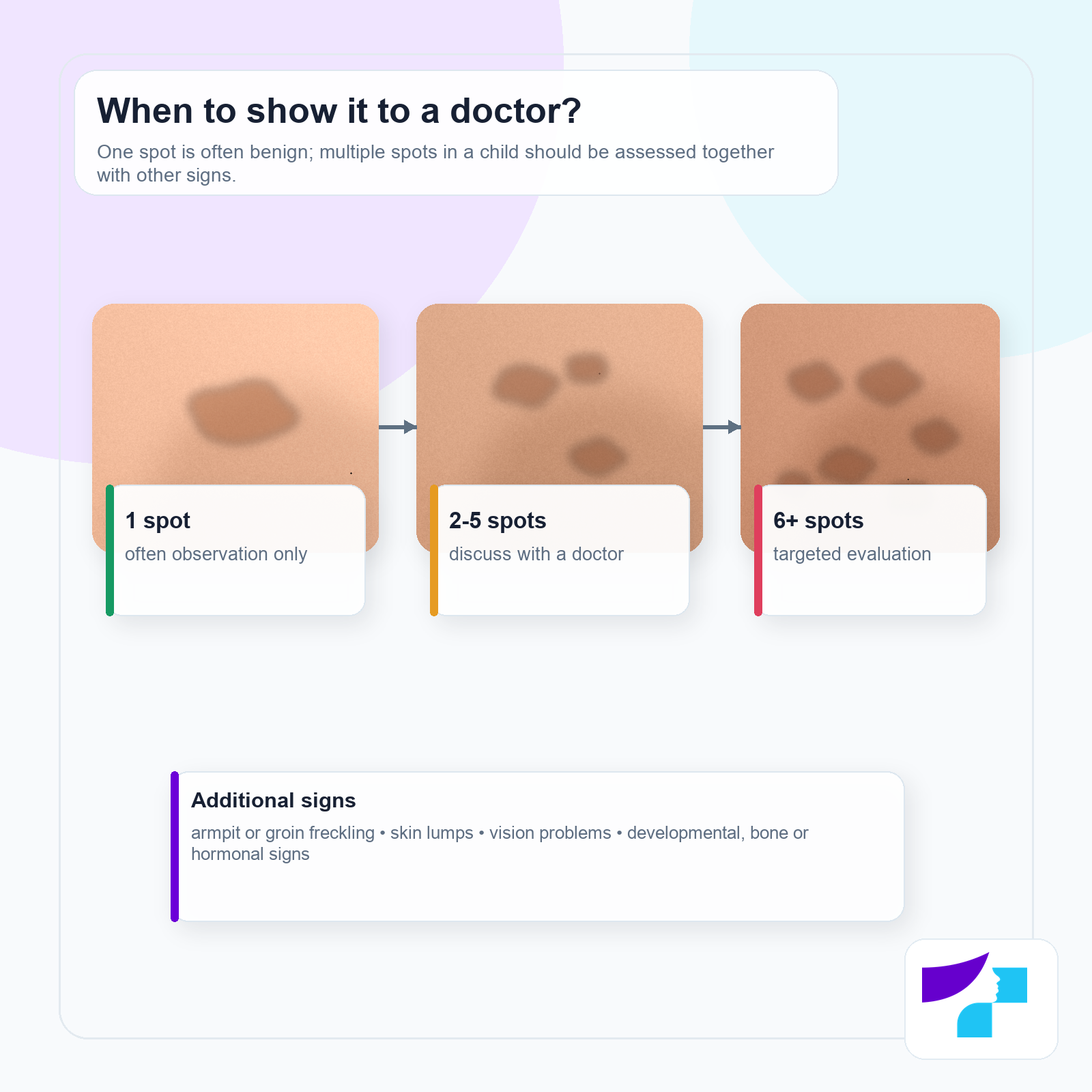
Important: a diagnosis is not made based only on the number of spots. For example, neurofibromatosis type 1 usually requires several diagnostic criteria or genetic confirmation.
Cafe-au-lait spots and neurofibromatosis
Neurofibromatosis type 1, or NF1, is a genetic condition that may affect the skin, nervous system, eyes, bones and development. Cafe-au-lait spots are one of the most common early skin signs of NF1, but they are not a diagnosis by themselves.
A doctor may consider NF1 if there are:
- six or more cafe-au-lait spots;
- spots that meet size criteria depending on the patient’s age;
- freckle-like pigmentation in the armpits or groin;
- two or more neurofibromas or one plexiform neurofibroma;
- optic pathway glioma;
- Lisch nodules in the iris or other characteristic eye signs;
- typical bone changes;
- a first-degree relative with NF1;
- a confirmed pathogenic variant in the NF1 gene.
If a child has multiple cafe-au-lait spots but no other signs, the doctor may recommend observation over time, an eye examination, genetic counselling or genetic testing. Some NF1 signs may appear later in childhood, so a single examination does not always fully rule out the diagnosis.
Other possible syndromes and conditions
Cafe-au-lait spots may also occur in other conditions. This is why the whole clinical picture matters.
| Condition | Typical features | What it means for the patient |
|---|---|---|
| Legius syndrome | Multiple cafe-au-lait spots, sometimes armpit or groin freckling, macrocephaly, learning or attention difficulties; without the typical NF1 tumour features | May resemble NF1 in early childhood; genetic testing may be needed |
| McCune-Albright syndrome | Large irregular pigmented patches, often along the midline; fibrous dysplasia of bone; hormonal problems such as early puberty | May require evaluation by an endocrinologist, orthopaedic specialist or other specialists |
| Noonan syndrome with lentigines | Multiple pigmented spots with heart, facial, growth or other features | Requires genetic and multidisciplinary evaluation |
| Isolated multiple cafe-au-lait spots | Multiple spots without other findings | May be benign, but children usually need observation |
How to distinguish a cafe-au-lait spot from a mole?
It is not always possible for a patient to distinguish them reliably by appearance, so any atypical or changing pigmented lesion should be assessed by a dermatologist.
| Feature | Cafe-au-lait spot | Mole, or melanocytic nevus |
|---|---|---|
| Surface | Flat and smooth | May be flat or raised |
| Colour | Usually evenly light-to-dark brown | May be brown, black, pinkish or uneven in colour |
| Sensation | Usually no symptoms | Usually no symptoms, but changes are important |
| Dermoscopy | Area of hyperpigmentation without a typical nevus structure | May show melanocytic nevus structures |
| Significance | Often benign; multiple spots may be a syndrome sign | Most are benign, but some need monitoring according to skin cancer risk criteria |
If a pigmented lesion becomes asymmetric, changes colour, grows rapidly, bleeds, ulcerates, becomes painful or raised, it should be shown to a dermatologist.
Differential diagnosis: what can it be confused with?
| Condition | How it may differ |
|---|---|
| Lentigo | Usually smaller and clearly pigmented; more often associated with sun exposure or ageing. |
| Becker nevus | More often appears during adolescence; may be larger, unevenly pigmented and hairy. |
| Congenital melanocytic nevus | May be a mole-type lesion with a different dermoscopic structure and different monitoring approach. |
| Post-inflammatory hyperpigmentation | Appears after eczema, trauma, burns, acne or another inflammatory process. |
| Dermal melanocytosis | Usually has a bluish-grey colour, often in the sacral or buttock area in infants. |
| Inflammatory or infectious skin disease | Usually has other signs such as itching, scaling, redness, peeling or rash. |
A cafe-au-lait spot is not the same as a sun or age spot
Patients sometimes think of a cafe-au-lait spot as just another “pigment spot”, but medically it is not the same as a sun spot, age spot or melasma.
Sun or age spots usually develop later in life and more often appear on sun-exposed areas such as the face, hands, décolletage or shoulders. Melasma is more often a symmetrical, diffuse facial hyperpigmentation that may be related to hormonal changes and sun exposure. Post-inflammatory hyperpigmentation appears after inflammation, trauma, acne, eczema or skin irritation.
A cafe-au-lait spot, by contrast, is often present at birth or appears in early childhood. It may occur anywhere on the body and is not a typical sign of sun damage. Sun protection cannot prevent the development of cafe-au-lait spots, although it remains important for overall skin health and for reducing other pigmentation problems.
When should you see a doctor about a cafe-au-lait spot?
You should see a doctor if:
- a child has multiple cafe-au-lait spots;
- the number of spots increases;
- the spots are large or there are six or more;
- there are freckle-like spots in the armpits or groin;
- there are lumps on or under the skin;
- there are vision problems, squinting or other eye symptoms;
- there are seizures, headaches or neurological symptoms;
- there is developmental delay, speech delay or learning difficulties;
- there are signs of early puberty;
- there is bone pain, deformity or frequent fractures;
- a pigmented lesion bleeds, ulcerates, hurts or rapidly changes colour or shape.
Urgent help is needed if seizures, sudden loss of vision, severe neurological symptoms, rapidly worsening severe pain or another acute condition occurs. In Latvia, call 113 or 112 in such cases.
How does a doctor diagnose a cafe-au-lait spot?
A cafe-au-lait spot is usually diagnosed clinically by examining the skin. A medical history and physical examination are often enough. The doctor may ask:
- when the spot first appeared;
- whether it is enlarging;
- whether new spots are appearing;
- whether similar spots, NF1 or other genetic conditions occur in the family;
- whether the child has developmental, vision, learning or neurological signs.
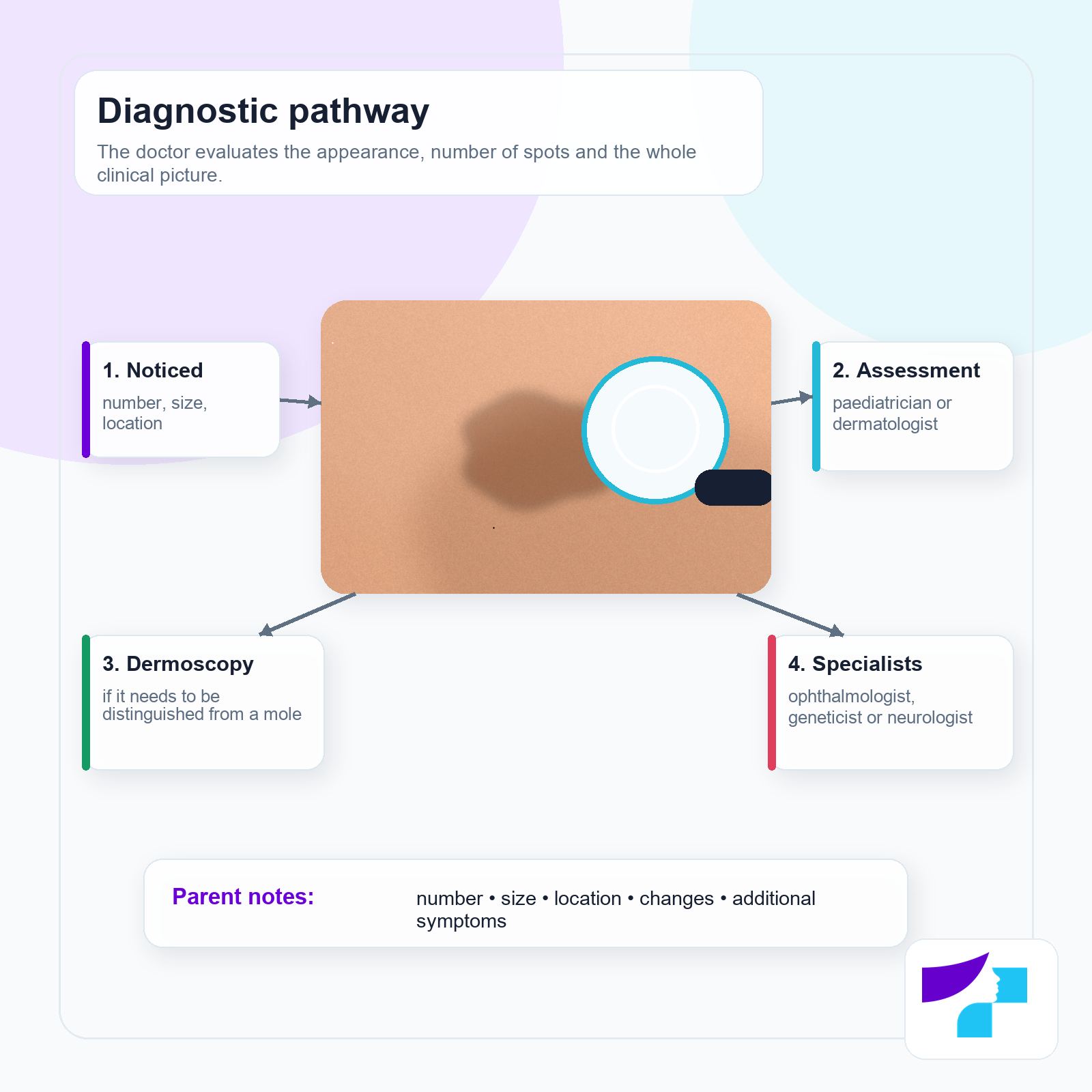
The assessment may include a full skin examination, counting and measuring the spots, dermoscopy if the lesion needs to be distinguished from a mole or another pigmented lesion, and involvement of a paediatrician, dermatologist, ophthalmologist, geneticist, neurologist, endocrinologist or orthopaedic specialist if needed.
A biopsy is usually not needed for a typical cafe-au-lait spot. It may be needed only if the lesion looks atypical and the doctor needs to rule out another diagnosis.
Do cafe-au-lait spots disappear or enlarge?
Many cafe-au-lait spots persist throughout life. In children, they may become more visible and enlarge because the skin and body are growing. This alone is not necessarily a concerning sign.
However, proportional growth should be distinguished from rapid change. If a spot suddenly changes colour, becomes raised, starts bleeding or ulcerating, or changes rapidly in appearance, this is no longer a typical cafe-au-lait scenario and a dermatologist should assess it.
Can cafe-au-lait spots be treated or removed?
Medical treatment for an isolated cafe-au-lait spot is usually not needed. If the spot is benign and causes no symptoms, explanation and observation are usually sufficient.
Laser treatment is sometimes considered for cosmetic reasons. Different pigment lasers may be used in dermatology, but results can vary:
- the spot may become lighter but not disappear completely;
- several procedures may be needed;
- the result depends on the type of spot, its location and skin tone;
- pigmentation may recur;
- side effects may include temporary or persistent hyperpigmentation, hypopigmentation, irritation or scarring.
Laser treatment should therefore not be seen as guaranteed “removal”. It should be discussed individually with a dermatologist.
Before laser treatment, it is advisable to:
- make sure the lesion is truly a typical cafe-au-lait spot;
- have a dermatologist’s assessment and, if necessary, dermoscopy;
- first evaluate a possible syndromic cause in children with multiple spots;
- discuss realistic results, recurrence risk and possible side effects;
- not delay medical evaluation simply because the spot is planned for cosmetic removal.
What should not be done to a pigmented spot?
You should not:
- try to bleach the spot with acids, lemon, unsuitable cosmetic products or unknown preparations;
- scratch, scrub or mechanically damage the spot;
- use prescription ointments without a doctor’s advice;
- assume that multiple spots in a child will “definitely go away” without evaluation;
- assume that one spot automatically means neurofibromatosis;
- delay a visit if there are also eye, nervous system, bone, developmental or hormonal signs.
How can parents monitor a child’s spots?
Parents do not need to make a medical diagnosis, but observation can help the doctor.
| What to monitor | How to record it |
|---|---|
| Number | How many spots are visible in total |
| Size | Approximate size in millimetres or centimetres |
| Location | For example, back, abdomen, leg, armpit |
| Changes | Whether new spots appear or existing spots change |
| Additional signs | Vision, development, learning, skin lumps, bone pain |
Photographs can help compare changes over time, but they should not be used for self-diagnosis. Ideally, take photos in similar lighting conditions, with a ruler next to the spot if the doctor asks you to monitor its size.
How to prepare for a doctor’s visit?
Before the visit, it is useful to prepare:
- when the first spot was noticed;
- whether the spots were present at birth;
- whether new spots have appeared;
- whether similar spots, NF1, tumours, early puberty or bone problems occur in the family;
- whether the child has vision, developmental, learning or neurological symptoms;
- previous photographs, if available;
- questions about whether a dermatologist, ophthalmologist or genetic specialist is needed.
Common myths about cafe-au-lait spots
| Myth | Fact |
|---|---|
| A cafe-au-lait spot always means neurofibromatosis. | No. One or a few spots are often benign. Multiple spots should be evaluated by a doctor. |
| One “coffee with milk” spot is dangerous. | Usually no, if it is typical and there are no other signs. |
| A cafe-au-lait spot is a mole. | Not exactly. It is a pigmentation patch, not a typical melanocytic mole. |
| It can easily become skin cancer. | A typical isolated cafe-au-lait spot is usually not a precancerous lesion. |
| The spot can safely be bleached at home. | No. Aggressive home remedies can damage the skin. |
| Laser always removes the spot completely. | No. Results vary and pigmentation may return. |
| A child with multiple spots will definitely have a serious disease. | Not always, but medical evaluation is needed. |
| The spots always disappear as the child grows. | They often persist, although their appearance may change. |
| An enlarging spot is always a bad sign. | In a child, the spot may grow with the body. Rapid or atypical changes should be assessed. |
| Sun exposure is the main cause of cafe-au-lait spots. | Usually no. They are related to a local increase in melanin, not simply tanning. |
What is well established in medicine and where is there still uncertainty?
Well established:
- cafe-au-lait spots are flat hyperpigmented skin patches;
- isolated spots are often benign;
- multiple spots may be associated with NF1 and other genetic conditions;
- NF1 diagnosis is based on a set of criteria, not just one spot;
- Legius syndrome may resemble NF1 because of pigmentation signs, but lacks the typical NF1 tumour manifestations;
- in McCune-Albright syndrome, pigmented patches may be more irregular and associated with bone or endocrine signs;
- laser treatment results are variable.
Areas with clinical uncertainty or individual variation:
- how early NF1 can be reliably distinguished from Legius syndrome in a child with multiple spots based only on clinical findings;
- which patient will have a stable and complete cosmetic result from laser treatment;
- how best to structure long-term monitoring for children with multiple spots but no other signs initially.
Current approaches:
- broader genetic testing when NF1, Legius syndrome and other RASopathy spectrum conditions need to be distinguished clinically;
- more precise monitoring of eye, skin and neurological signs in children with suspected NF1;
- ongoing development of laser technologies for cosmetic pigment reduction, while maintaining caution about variable results.
Summary
A cafe-au-lait spot, or “coffee with milk” spot, is usually a flat, evenly brown skin patch. One or a few such spots are often benign and do not require treatment. More attention is needed when there are multiple spots, when they are large, or when they appear in a child together with armpit or groin freckling, skin lumps, vision, developmental, bone, hormonal or neurological signs.
The most important point is not to diagnose the condition yourself, but to know when medical assessment is needed. A dermatologist, paediatrician or genetic specialist can assess whether the spots are simply a benign skin feature or part of a broader medical condition.
Medical disclaimer
This information is intended for educational purposes only and does not replace medical consultation, diagnosis or treatment. If a child or adult has multiple cafe-au-lait spots, if the spots change, if new lesions appear, or if there are signs involving the eyes, nervous system, bones, hormones, development or other general health concerns, it is recommended to consult the clinic’s dermatologist for assessment. Do not try to bleach, damage or treat pigmented spots with unverified products or prescription medicines without medical advice.
References and authoritative sources
- DermNet: clinical information on cafe-au-lait macules, appearance, associated conditions and treatment options
- NCBI Bookshelf, StatPearls: Cafe Au Lait Macules overview of epidemiology, diagnosis and associated syndromes
- GeneReviews: Neurofibromatosis 1 diagnostic criteria, clinical features and monitoring
- GeneReviews: Legius Syndrome clinical features and differentiation from neurofibromatosis type 1
- GeneReviews: Fibrous Dysplasia / McCune-Albright Syndrome and associated skin, bone and endocrine features
- Cleveland Clinic: cafe-au-lait spot causes, diagnosis and treatment options in patient-friendly language
- MSD Manual Professional Edition: clinical signs and diagnosis of neurofibromatosis
- Centre for Disease Prevention and Control of Latvia: public health information resource
- Ministry of Health of the Republic of Latvia: national health care information
