Lentigo simplex, or simple lentigo, is a benign pigmented spot on the skin or mucous membrane. It is usually flat, round or oval, and light brown, dark brown or sometimes almost black in colour. It most often appears in childhood or young adulthood and, unlike sun-related pigment spots, is not directly caused by sun exposure.
In most cases, lentigo simplex is not dangerous and is not skin cancer. However, any new, changing, asymmetrical, multicoloured, itchy, bleeding or rapidly growing pigmented lesion should be examined by a dermatologist, because melanoma and other skin cancers can sometimes resemble benign pigmented spots.
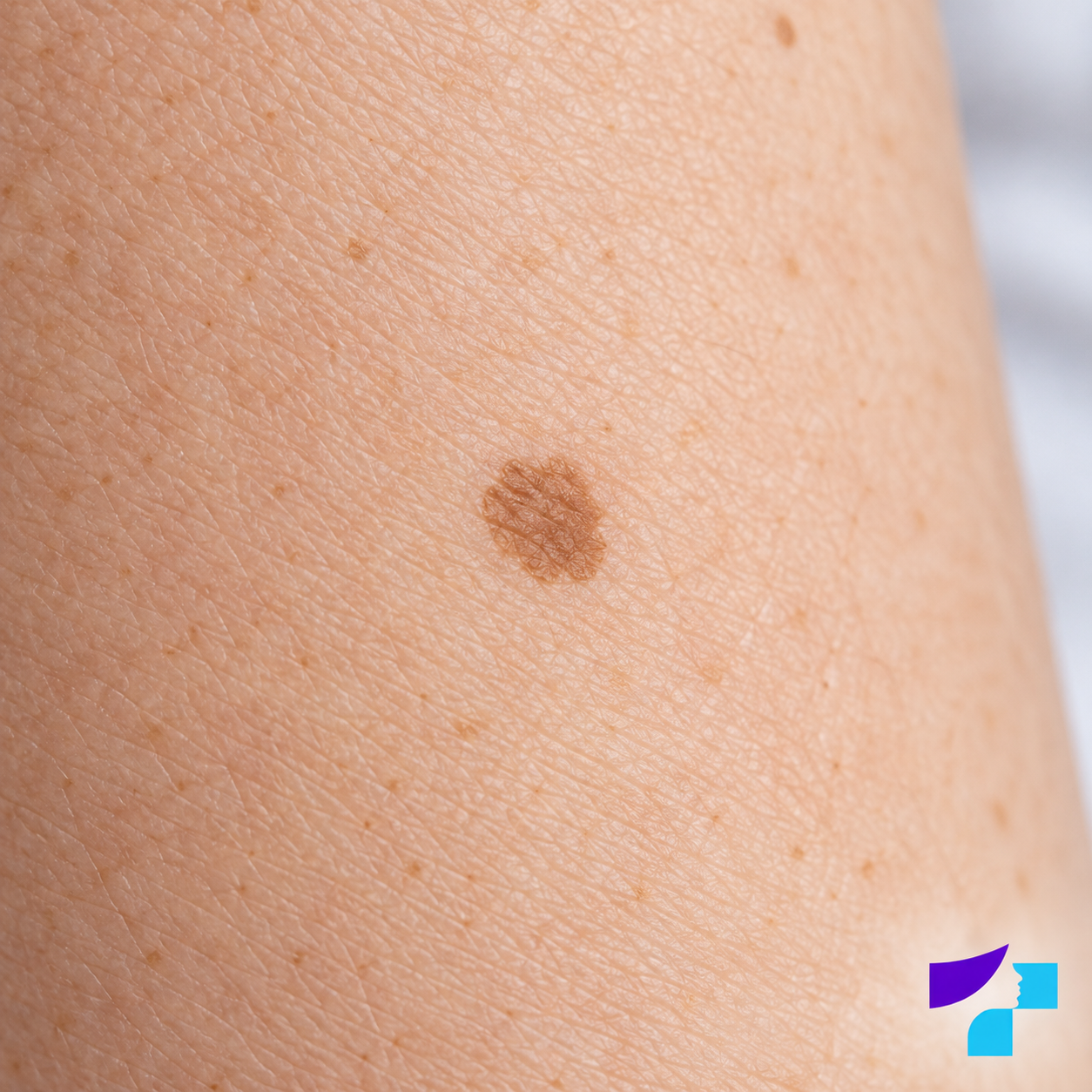
The image shows lentigo simplex, or simple lentigo - a flat, benign, evenly pigmented spot on the skin.
What is lentigo simplex?
Lentigo simplex is one type of lentigo. In dermatology, the term “lentigo” refers to a clearly defined pigmented spot in which there is an increased amount of melanin and increased melanocyte activity. Melanocytes are cells that produce melanin - the pigment that gives colour to the skin, hair and eyes.
Lentigo simplex is also called simple lentigo or juvenile lentigo, because it may appear in childhood or young adulthood. In everyday language, it may be described as a common pigmented spot, but medically the term “pigmented spot” is broader: it may refer to lentigo simplex, solar lentigo, melasma, post-inflammatory hyperpigmentation or another pigmentation change.
What does lentigo simplex look like?
A typical lentigo simplex is:
- a flat spot, or macule;
- round or oval;
- usually a few millimetres to about 1-1.5 cm in diameter;
- uniformly light brown, dark brown or black;
- clearly demarcated, with smooth or slightly irregular borders;
- usually smooth on the surface, although it may sometimes appear slightly dry;
- not painful, itchy or bleeding.
Lentigo simplex may be single or multiple. It can occur on the body, limbs, face, lips, oral mucosa, other mucous membranes and very rarely on the conjunctiva of the eye. If there are very many pigmented spots, if they appear suddenly or if they are associated with other symptoms, assessment by a dermatologist is needed, because in rare cases multiple lentigines may be associated with inherited syndromes.
Why does lentigo simplex develop?
The exact cause of lentigo simplex is not always known. It develops because of local changes in pigment cells and melanin, but it is usually not directly related to sun exposure. This is important because many people call any brown spot a “sun spot”, although lentigo simplex and solar lentigo are not the same condition.
Lentigo simplex may also appear in people who do not have obvious sun-damaged skin. Some sources note that multiple lentigines may be seen more often in people with fair skin or red hair, but a single stable lentigo simplex usually does not indicate an internal disease. Broader evaluation is needed if there are many spots, if they appear at an early age in unusual locations or if they are associated with other signs, such as developmental, gastrointestinal, endocrine, eye or heart problems.
Is lentigo simplex dangerous?
Lentigo simplex itself is benign and is not skin cancer. It usually does not cause pain, inflammation or general health problems. However, self-diagnosis should be avoided, because benign pigmented spots, moles, solar lentigo, lentigo maligna and melanoma may overlap in appearance.
The safest principle is simple: a stable, unchanged, uniformly coloured and symmetrical spot is usually less concerning, but a new or changing spot should be shown to a doctor.
Lentigo simplex in children and adults
Lentigo simplex may be present from birth, but more often it appears in childhood or young adulthood. In a child, one or a few stable pigmented spots are usually not a reason for alarm, but they should be shown to a dermatologist if they change, become very dark, bleed, grow rapidly or appear in large numbers.
In adults, a new pigmented lesion should always be assessed more carefully. Not every new brown spot means cancer, but in adulthood it is especially important to rule out melanoma, lentigo maligna, pigmented actinic keratosis or other lesions.
Where on the body can it appear?
Lentigo simplex can appear anywhere on the skin or mucous membranes. It is not limited to sun-exposed areas. It may occur on the trunk, arms, legs, face, lips, oral mucosa or other areas. Medical sources also mention the conjunctiva as a possible but rare location - the thin mucous membrane that covers the front surface of the eye and the inner surface of the eyelids.
If a pigmented spot is located on the lips, inside the mouth, in the genital area, on the palms, soles or under a nail, it is advisable to have it examined by a dermatologist, because visual assessment in these areas can be more difficult.
Lentigo simplex and freckles - what is the difference?
Freckles, or ephelides, are usually small, light brown spots that become more noticeable with sun exposure and often fade in winter. They are more common in people with fair skin, light hair or red hair.
Lentigo simplex is usually darker, less numerous, more clearly defined and does not change as strongly with the seasons. It is not a classic sun-induced spot.
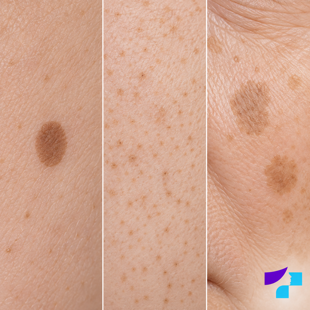
The image compares different benign pigmentation patterns: lentigo simplex, freckles and solar lentigo, also known as sun-related pigmented spots.
| Feature | Lentigo simplex | Freckles |
|---|---|---|
| Relationship with sun | Usually not directly related | Become more pronounced in the sun |
| Seasonal changes | Usually more stable | Often darker in summer and lighter in winter |
| Colour | Light brown to dark brown/black | Usually light brown |
| Number | May be single or multiple | Often numerous |
| Age | Childhood, young adulthood or later | Often childhood |
Lentigo simplex and solar lentigo - what is the difference?
Solar lentigo is a pigmented spot that develops as a result of long-term ultraviolet radiation exposure. It is often called an age spot or sun spot, and it is commonly discussed in the context of skin ageing. It most often appears on the face, backs of the hands, shoulders, décolletage and lower legs.
Lentigo simplex usually appears at a younger age and is not directly caused by UV radiation. In practice, however, these types of spots can sometimes be difficult to distinguish by appearance alone, so a dermatologist may use dermoscopy.
| Feature | Lentigo simplex | Solar lentigo |
|---|---|---|
| Main factor | Unclear; not directly sun-induced | Chronic UV exposure |
| More common age | Childhood, young adulthood | Middle and older age |
| Typical location | Anywhere on the skin or mucous membranes | Sun-exposed areas |
| Prevention | No specific prevention | Sun protection reduces the risk of new spots |
Lentigo simplex and a mole - what is the difference?
A mole, or melanocytic naevus, is a collection of melanocytes. It may be flat, raised, light, dark, round or irregular. Lentigo simplex is a flat pigmented spot, not a typical mole formed by nests of melanocytes.
For patients, the most important practical point is not to determine the exact diagnosis themselves, but to notice changes. If a lesion grows, changes, becomes multicoloured, bleeds or looks different from other skin lesions, a dermatologist consultation and often a mole check are needed.
Pigmented spot or melanoma - when should you be concerned?
Melanoma is a malignant skin tumour that may be brown, black, reddish, pink, bluish or multicoloured. It may develop from an existing mole or appear as a new lesion. Some melanomas may initially look like an apparently harmless pigmented spot.
You should see a dermatologist if:
- the spot grows rapidly;
- its colour, shape or surface changes;
- it becomes asymmetrical;
- the borders become blurred, jagged or uneven;
- several colours appear within one lesion;
- it starts itching, hurting, bleeding or forming crusts;
- it is new and looks different from other skin lesions;
- it is located under a nail, on the palm, sole, lip or mucous membrane and changes.
ABCDE signs in simple language
The ABCDE principle helps identify lesions that should be shown to a dermatologist:
- A - asymmetry: one half does not look like the other;
- B - borders: the edges are jagged, blurred or poorly defined;
- C - colour: there are several colours in one spot or unusually dark areas;
- D - diameter: the lesion is larger than about 6 mm, although melanoma may also be smaller;
- E - evolution: the lesion changes in size, shape, colour, height or symptoms.
The “ugly duckling” sign is also important: one lesion looks different from the person’s other skin lesions.
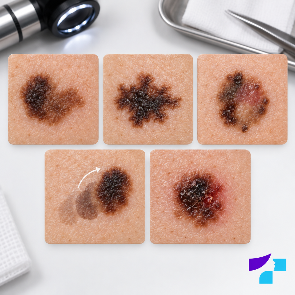
The image shows examples of features that should prompt a dermatologist check: asymmetry, irregular borders, multiple colours, visible change or crusting.
How does a dermatologist diagnose a pigmented spot?
The dermatologist assesses the appearance, size, colour, symmetry, borders and changes over time, as well as the patient’s age, skin type, sun exposure, family history, previous skin tumours and other risk factors.
The examination usually includes visual skin assessment and dermoscopy. Dermoscopy is an examination using a special magnifying device that allows the doctor to see pigment structures more deeply and precisely than with the naked eye. A typical dermoscopic feature of lentigo simplex may be a symmetrical, uniform pigment network that ends fairly abruptly at the edge of the spot. This helps the doctor distinguish lentigo simplex from some melanocytic naevi, in which pigment may fade more gradually towards the edge. If there are multiple lesions or an increased melanoma risk, skin lesion mapping and follow-up monitoring may be recommended.
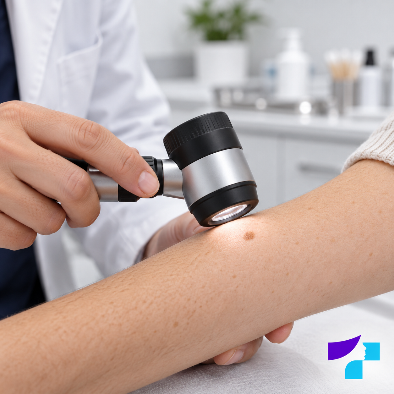
The image shows a pigmented spot being examined with a dermoscope - a magnifying device that helps the dermatologist assess the structure of a pigmented lesion more accurately.
When is biopsy or histology needed?
If the dermatologist is uncertain after examination or dermoscopy, the lesion may be excised or a tissue sample may be taken for histological examination. Histology means examining tissue under a microscope, and it is the most reliable way to distinguish a benign lesion from a malignant one.
Microscopically, lentigo is usually characterised by increased melanin and an increased number of melanocytes in the basal layer of the epidermis. Unlike many melanocytic naevi, melanocytes usually do not form pronounced “nests”. Patients do not need to remember this detail, but it helps explain why histology can clarify the diagnosis if the clinical assessment is not completely clear.
A suspicious pigmented lesion should not first be cosmetically lightened, burned or treated with laser without dermatologist assessment, because this may make diagnosis more difficult and delay appropriate treatment.
Does lentigo simplex need treatment?
If lentigo simplex is stable, benign and does not cause cosmetic concern, treatment is usually not necessary. It can simply be monitored. Some spots may become lighter over time, while others persist for years.
Treatment or removal may be considered when the diagnosis is clear and the spot causes cosmetic concern or is located in an area where it is frequently traumatised. Before any cosmetic procedure, it is important to make sure that the lesion is not suspicious.
Options for removing pigmented spots
Depending on the type and location of the lesion, a dermatologist may consider:
- laser therapy with pigment lasers;
- cryotherapy, or freezing;
- chemical peeling;
- topical pigment-reducing treatments, such as retinoids, azelaic acid or hydroquinone, if the doctor considers them suitable;
- surgical excision;
- observation without a procedure.
Not all methods are suitable for all pigmented spots. Laser therapy may be appropriate for some benign pigment lesions, but it is not the right choice for a suspicious lesion that needs histological examination. Cryotherapy, peels, laser therapy and active lightening agents may leave lighter spots, or hypopigmentation, darker spots, or hyperpigmentation, irritation or scars, especially in darker skin types. Therefore, procedures should be planned individually.
Which diagnoses can resemble lentigo simplex?
The differential diagnosis of lentigo simplex is broad. Similar lesions may include freckles, solar lentigo, moles, café-au-lait spots, melasma, post-inflammatory hyperpigmentation, seborrhoeic keratosis, nevus spilus, pigmented basal cell carcinoma, lentigo maligna and melanoma.
If the spot is not only pigmented but also scaly, itchy or inflamed, the doctor may also consider other skin diseases, such as psoriasis, eczema or an allergic skin reaction.
Some of these lesions are completely benign, while others require faster diagnosis or treatment. This is why the main safety principle of the article is not to call every brown spot lentigo simplex by yourself if it is new, unusual or changing.
What should you not do yourself?
You should not cut, burn, scratch, traumatise or aggressively lighten a pigmented spot yourself. It is not safe to use acids, “wart solutions”, folk remedies or online lightening products on a suspicious pigmented lesion. If the spot is melanoma or another type of skin tumour, such actions may delay diagnosis.
Lightening creams are also not a universal treatment for lentigo simplex. Some products may irritate the skin, cause post-inflammatory hyperpigmentation or mask changes. Dermatologist assessment is recommended before active pigment correction.
When should you see a dermatologist?
A dermatologist consultation is recommended if:
- a pigmented spot is new in adulthood;
- it changes;
- it looks different from other lesions;
- there is a personal or family history of melanoma;
- there are many moles or atypical moles;
- the lesion is on the lips, mucous membrane, palms, soles or under a nail;
- the person wants cosmetic removal of the pigmented spot.
When should the visit not be delayed?
The visit should not be delayed if a pigmented lesion bleeds without injury, grows rapidly, hurts, itches, ulcerates, forms crusts, becomes multicoloured, markedly asymmetrical or rapidly changes shape. A new suspicious lesion after the age of 30 should be assessed especially carefully.
How to prepare for a dermatologist visit?
Before the visit, it is useful to write down:
- when the spot appeared;
- whether it has changed;
- whether there is itching, pain, bleeding or crusting;
- whether the spot has been traumatised or treated with creams;
- whether there is a family history of melanoma or skin cancer;
- whether the patient has previously had skin tumours;
- whether there have been frequent sunburns or tanning bed use.
Concealing make-up should not be applied to the area being examined before the visit. If there are older photographs in which the spot is visible, they may help assess changes over time.
Common myths about lentigo simplex
| Myth | Fact |
|---|---|
| Lentigo simplex is melanoma | No, it is a benign pigmented spot, but suspicious lesions should be examined |
| All pigmented spots are harmless | Many are benign, but some skin tumours may resemble them |
| Lentigo simplex always develops from the sun | It is usually not directly caused by sun exposure |
| A pigmented spot can be safely removed at home | Suspicious pigmented lesions should not be burned or cut at home |
| Laser is always the best method | A suspicious lesion first needs diagnosis, not cosmetic treatment |
| If the spot does not itch, it cannot be dangerous | Many suspicious lesions cause no symptoms at first |
| If the spot is smaller than 6 mm, it is definitely not melanoma | Melanoma can also be smaller, especially at an early stage |
| A child’s pigmented spot is always dangerous | It is often benign, but changing or numerous spots should be checked |
| If a doctor diagnoses lentigo simplex, follow-up is never needed | Observation is usually enough, but follow-up may be needed if there are many spots or if the lesion changes |
Summary
Lentigo simplex, or simple lentigo, is usually a benign, flat, uniformly pigmented spot on the skin or mucous membrane. It often appears in childhood or young adulthood and is not directly related to sun exposure. The main practical message for patients is that lentigo simplex is usually not dangerous, but a pigmented lesion should not be self-diagnosed based on an internet description. If the spot is new, changing, bleeding, itchy, painful, asymmetrical or multicoloured, it should be examined by a dermatologist.
Medical disclaimer
This information is for educational purposes only and does not replace a dermatologist consultation, diagnosis or treatment. Accurate assessment of pigmented skin lesions depends on the appearance of the lesion, changes over time, dermoscopic findings and patient risk factors. If a pigmented spot changes rapidly, bleeds, hurts, itches, becomes asymmetrical, multicoloured or appears as a new suspicious lesion, you should see a dermatologist. Do not attempt to cut, burn or aggressively lighten suspicious pigmented lesions yourself.
References and authoritative sources
- DermNet NZ: medical information about lentigo simplex
- DermNet NZ: overview of lentigo and its types
- DermNet NZ: solar lentigo and sun-related pigmented spots
- Skinsight: patient information about lentigo simplex
- Primary Care Dermatology Society: clinical guidance on lentigo
- Dr. Magnus Lynch: differential diagnosis of lentigo simplex
- American Academy of Dermatology: ABCDE signs of melanoma
- Skin Cancer Foundation: warning signs of melanoma
- MSD Manual Professional Edition: hyperpigmentation disorders
- MSD Manual Professional Edition: melanoma
- NCBI MeSH: medical terminology for lentigo
- PubMed: biomedical publications database
- Centre for Disease Prevention and Control of Latvia: public health information
- Ministry of Health of the Republic of Latvia: health sector information
