A granuloma is a localized collection of inflammatory cells in the body. It is not one specific disease, but an immune system reaction to an irritant that the body cannot easily destroy or remove. A granuloma can form around an infection, a foreign body, chronic inflammation or in certain immune system diseases.
Short answer: a granuloma itself is usually not cancer, but its significance depends on its location, cause, symptoms and test results. In one situation, it may be an old, harmless finding on a lung X-ray; in another, it may be a sign of inflammation around a tooth root or an indication of a condition that requires treatment.
What is a granuloma?
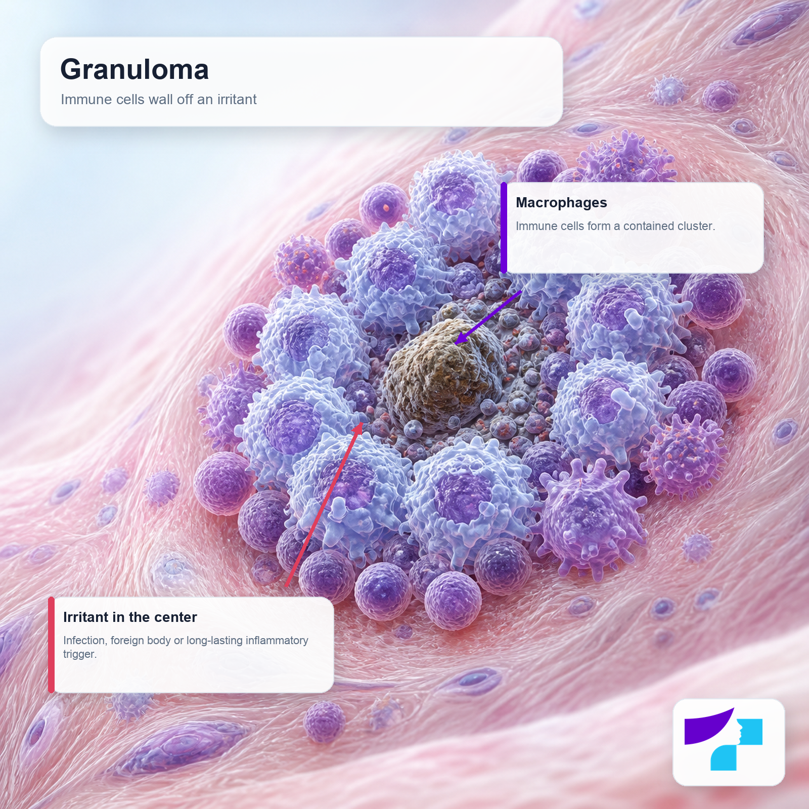
A granuloma is a small, organized group of immune cells that forms at the site of chronic inflammation. The main cells in a granuloma are macrophages - immune cells that usually help break down microbes, damaged cells and foreign substances.
If macrophages cannot fully destroy or remove the irritant, the body tries to wall it off. This creates granulomatous inflammation. The process can be compared to a protective barrier: the body attempts to “lock” the problem in one place so it does not spread further.
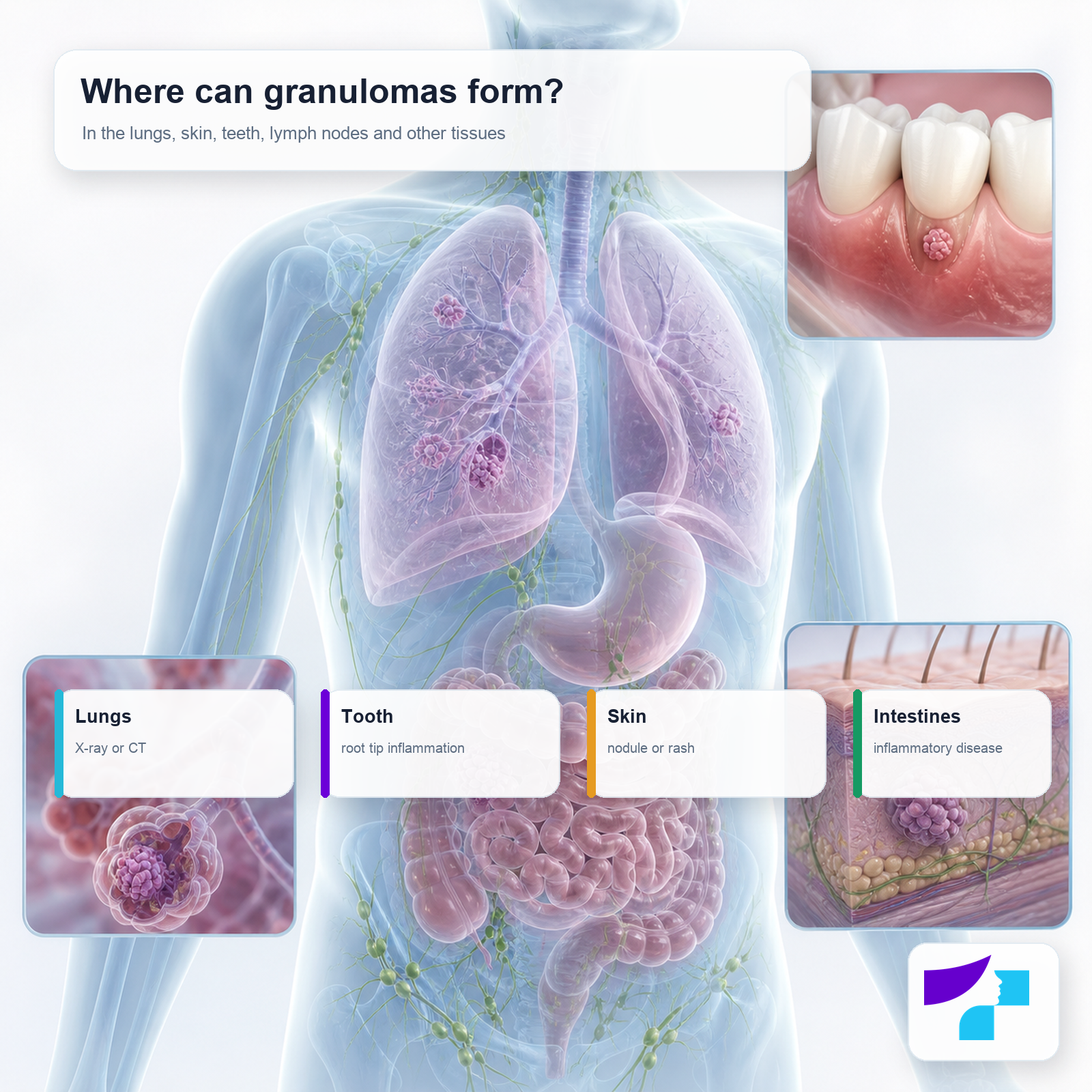
Granulomas can occur:
- in the lungs;
- in the skin;
- in lymph nodes;
- in the liver;
- in the intestines;
- in the mouth;
- near the tip of a tooth root;
- after surgery, trauma, injection or the entry of a foreign body into tissue.
Is a granuloma a disease or a diagnosis?
The word “granuloma” describes a type of tissue reaction rather than always naming a specific disease. Therefore, the most important question is not only “do I have a granuloma?”, but “why did it form?”.
For example:
- a dental granuloma is usually related to chronic inflammation near a tooth root;
- a lung granuloma may occur after a previous infection, sarcoidosis or another condition;
- granuloma annulare is a benign, non-contagious skin condition;
- a pyogenic granuloma is not actually a classic granuloma, but a benign vascular growth;
- a foreign body granuloma forms around material that the body cannot break down.
Why do granulomas form?
A granuloma forms when there is long-lasting irritation or an inflammatory trigger in the body. This may be infectious or non-infectious.
| Cause | Examples | What this means for the patient |
|---|---|---|
| Infection | Tuberculosis, nontuberculous mycobacteria, fungal infections | The exact cause should be identified and treated specifically |
| Foreign body | Suture, splinter, implant, filler, tattoo pigment | The reaction may persist while the material remains in the tissue |
| Chronic inflammation | Sarcoidosis, Crohn’s disease, rheumatologic diseases | The underlying condition usually needs to be treated |
| Dental infection | Damaged dental pulp, infection in the root canal | Treatment by a dentist or endodontist is required |
| Skin condition | Granuloma annulare, foreign body reaction, pyogenic granuloma | A dermatologist’s assessment is needed if the diagnosis is unclear |
Is a granuloma dangerous?
A granuloma is not always dangerous. Some granulomas are incidental findings on investigations and do not cause health problems. For example, a calcified granuloma in the lung often indicates an old, contained inflammatory process.
However, a granuloma can also be a sign of an infection, chronic inflammatory disease, dental infection or another condition. For this reason, the significance of a granuloma is determined by a doctor, taking into account:
- where it is located;
- whether there are symptoms;
- whether it changes over time;
- whether there are signs of infection or inflammation;
- what X-ray, CT, biopsy or blood test results show;
- the patient’s age, medical history and risk factors.
Is a granuloma cancer?
A granuloma is usually not cancer. It is an inflammatory reaction, not a malignant tumor. However, in some cases a granuloma or a nodule may look similar to another type of growth, including a tumor, on imaging or examination. Therefore, a doctor may recommend additional tests such as repeat imaging, biopsy or histological examination.
Medical assessment is especially important if the lesion:
- grows rapidly;
- bleeds;
- ulcerates;
- changes color or shape;
- becomes painful;
- is associated with weight loss, fever, night sweats or shortness of breath.
Symptoms of a granuloma
Many granulomas cause no symptoms. Symptoms depend on the location and cause of the granuloma.
| Location | Possible symptoms |
|---|---|
| Lungs | Persistent cough, shortness of breath, chest pain, fever, night sweats |
| Tooth | Pain when biting, sensitivity, gum swelling, fistula, bad taste in the mouth |
| Skin | Nodule, raised rash, ring-shaped patch, redness, itching, bleeding |
| Lymph nodes | Enlarged or tender lymph nodes |
| Intestines | Abdominal pain, diarrhea, weight loss, blood in stool |
| Eyes or nervous system | Vision problems, headaches or neurological signs in certain diseases |
Main types of granulomas
| Type of granuloma | Where it occurs | Typical features |
|---|---|---|
| Dental or periapical granuloma | Near the tip of a tooth root | Chronic inflammation, often visible on X-ray |
| Lung granuloma | In the lungs | May occur after infection, sarcoidosis or another inflammatory process |
| Calcified granuloma | Often in the lungs or lymph nodes | Usually a sign of an old, contained process |
| Skin granuloma | In the skin | Nodule, plaque or rash; must be distinguished from other skin conditions |
| Granuloma annulare | In the skin | Benign, non-contagious, often ring-shaped skin condition |
| Pyogenic granuloma | On the skin or mucous membrane | Benign vascular growth that often bleeds |
| Foreign body granuloma | Around foreign material in tissue | May occur after sutures, injections, implants or splinters |
| Granulomas in systemic diseases | Lungs, lymph nodes, intestines, skin and other sites | May be part of sarcoidosis, Crohn’s disease or rheumatologic diseases |
Dental granuloma or periapical granuloma
A dental granuloma usually means chronic inflammation near the tip of a tooth root. Medically, it is often called a periapical granuloma. It most commonly develops when the dental pulp is damaged by tooth decay, trauma or previous unsuccessful root canal treatment.
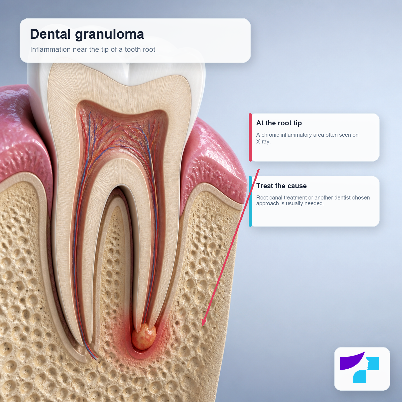
A dental granuloma may be painless and detected only on an X-ray. However, it can cause:
- pain when biting;
- tooth sensitivity;
- gum swelling;
- a fistula, or a small opening through which pus may drain;
- an unpleasant taste in the mouth;
- an acute flare-up with pain and swelling.
A dental granuloma usually does not go away on its own if the source of infection remains in the root canal. Antibiotics alone usually do not resolve the cause of chronic inflammation. Treatment may include root canal treatment, repeat endodontic treatment, root-end surgery or tooth extraction if the tooth cannot be saved.
Lung granuloma
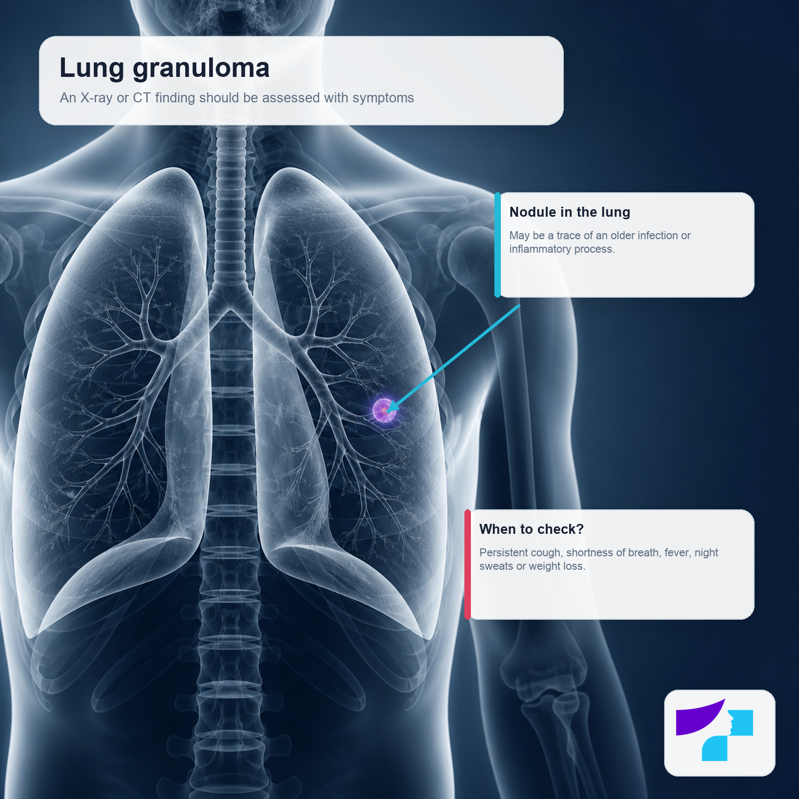
A lung granuloma is often found on X-ray or CT as a nodule or small localized change. It may be associated with:
- a previous infection;
- tuberculosis;
- nontuberculous mycobacteria;
- fungal infections;
- sarcoidosis;
- occupational exposure or inhaled irritants;
- less commonly, other inflammatory or immune system diseases.
A lung granuloma does not always mean tuberculosis or cancer. However, a doctor will assess whether observation, additional tests or treatment are needed. It is especially important to evaluate a granuloma if there is persistent cough, shortness of breath, fever, night sweats, weight loss or blood in sputum.
What does a calcified granuloma mean?
A calcified granuloma means that calcium deposits are present in the granuloma. This often indicates an old, contained process that is no longer active. Such granulomas may remain for years and be repeatedly described on imaging reports.
However, a calcified granuloma should still be assessed in context. The doctor considers:
- whether it is stable over time;
- whether previous imaging is available for comparison;
- whether the patient has symptoms;
- whether there are risk factors for tuberculosis, fungal infection or other diseases;
- whether follow-up imaging is needed.
Skin granuloma
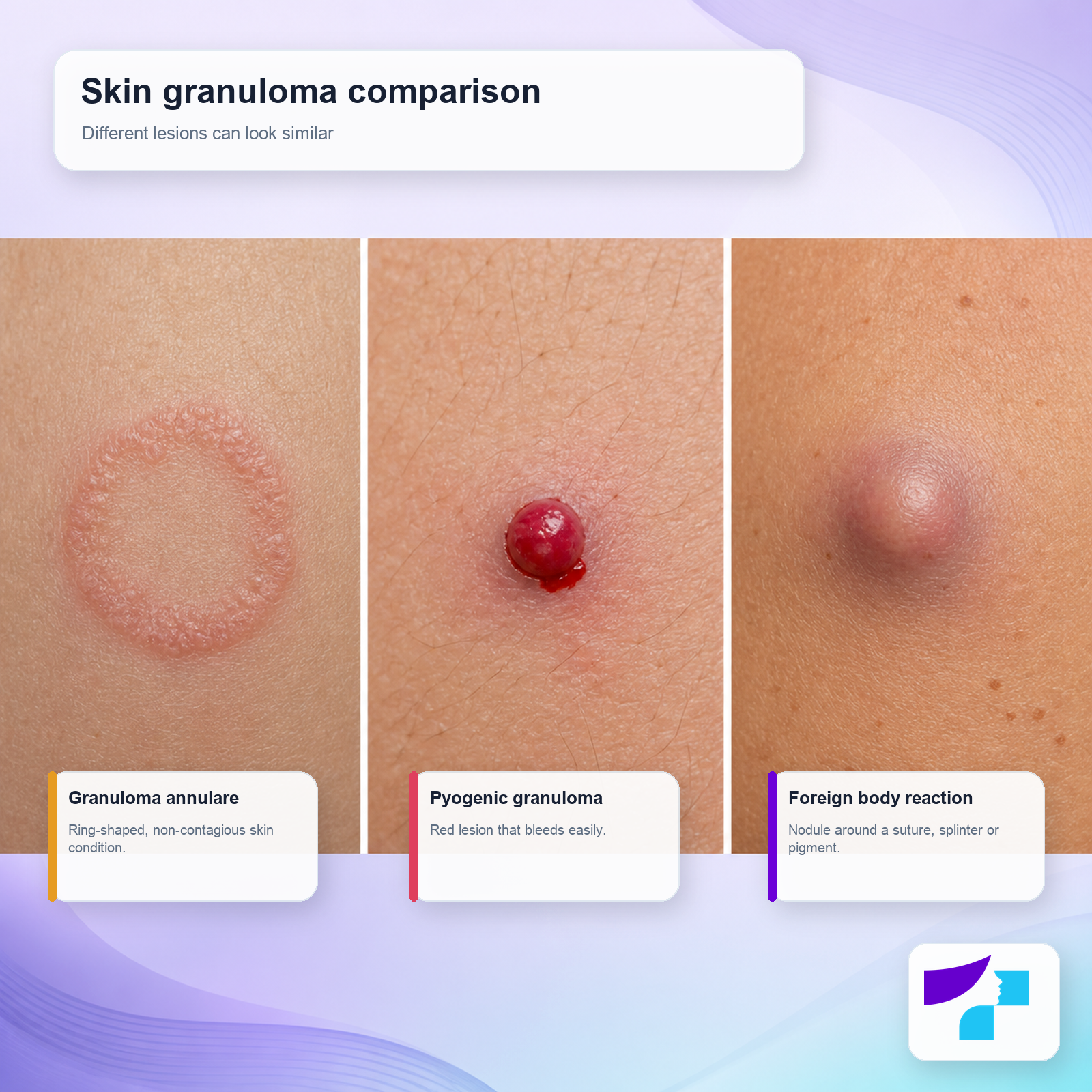
A skin granuloma may look like a firm nodule, raised area, red plaque, ring-shaped rash or a lesion that bleeds easily. Different skin lesions can look similar, so an accurate diagnosis often requires assessment by a dermatologist.
For a skin granuloma, a doctor may use:
- clinical examination;
- dermoscopy;
- skin scraping if a fungal infection needs to be ruled out;
- biopsy if the diagnosis is unclear or the lesion looks atypical.
Granuloma annulare
Granuloma annulare is a benign, non-contagious skin condition. It often appears as a ring-shaped rash or small raised bumps arranged in a circle. It is most commonly seen on the hands, feet, forearms, elbows, ankles or other areas of the limbs.
It is not an infection and it is not cancer. In many patients it causes no pain and clears on its own, although this may take months or even years. In some people, the rash may recur.
Main forms:
- localized granuloma annulare - one or a few ring-shaped lesions;
- generalized granuloma annulare - lesions over wider areas of the body;
- subcutaneous form - firm lumps under the skin, more common in children;
- perforating form - a rarer form with superficial skin changes;
- patch form - flatter, reddish or brownish skin areas.
Treatment is not always needed. If the lesions are widespread, bothersome or persistent, a dermatologist may consider topical corticosteroids, injections, cryotherapy, phototherapy or other methods. Treatment results can vary.
Pyogenic granuloma
Pyogenic granuloma is a misleading name. It is not a pus-forming infection and it is not a classic granuloma. Medically, it is a benign proliferation of capillary blood vessels, or a vascular growth.
A pyogenic granuloma usually appears as a red, moist, rapidly growing bump that bleeds easily. It can occur on the skin or mucous membranes, including in the mouth. It may be promoted by trauma, chronic irritation, pregnancy, certain medicines or poor oral hygiene when the lesion is on the gums.
Because a pyogenic granuloma can bleed and sometimes resemble other skin lesions, it should be shown to a dermatologist, dentist or another appropriate specialist. Removal and histological examination may sometimes be needed.
Foreign body granuloma
A foreign body granuloma forms around material that the body cannot fully break down. This may include:
- a surgical suture;
- a splinter;
- an implant;
- injected filler;
- tattoo pigment;
- a fragment of glass, metal or another material;
- a reaction after an injection or procedure.
This type of granuloma may be a firm nodule, tender thickening or a zone of chronic inflammation. Treatment depends on the symptoms and cause. Sometimes observation is enough, while in other cases removal of the foreign body, anti-inflammatory therapy or surgery may be needed.
What does a granuloma mean in a biopsy?
A biopsy is the removal of a tissue sample for examination under a microscope. If a histology report mentions a granuloma or granulomatous inflammation, it means that the pathologist has seen a specific type of inflammatory reaction in the tissue.
Histology may describe several types of granulomas:
- caseating granuloma - with tissue necrosis in the center; more often suggests certain infections, such as tuberculosis, but should not be interpreted in isolation;
- non-caseating granuloma - without this type of necrosis; may occur in sarcoidosis, foreign body reactions and other conditions;
- necrobiotic granuloma - associated with collagen damage, for example in granuloma annulare;
- suppurative granuloma - with a pus-forming inflammatory component; may occur in certain infections;
- foreign body granuloma - with a reaction around material the body cannot break down.
Important: the granuloma type helps the doctor consider the possible cause, but it does not always determine the final diagnosis by itself.
How does a doctor diagnose a granuloma?
Diagnosis depends on where the granuloma is located and what symptoms are present.

| Test or examination | When it is used |
|---|---|
| Clinical examination | To assess skin, mouth, lymph nodes or visible lesions |
| Dental X-ray | If a tooth root granuloma is suspected |
| Chest X-ray | If a granuloma or nodule is seen in the lungs |
| Computed tomography | For more detailed assessment of the lungs, lymph nodes or organs |
| Ultrasound | For superficial nodules or abdominal organs |
| Dermoscopy | To assess skin lesions |
| Blood tests | To assess infection, inflammation or organ function |
| Biopsy | If the diagnosis is unclear or other conditions must be ruled out |
| Microbiological tests | If bacteria, mycobacteria or fungi need to be looked for |
When is a biopsy needed?
A biopsy may be needed if:
- the diagnosis is unclear;
- the lesion is growing rapidly;
- a skin lesion bleeds, ulcerates or looks atypical;
- an imaging finding cannot be confidently explained;
- infection, tumor or another disease must be ruled out;
- treatment does not produce the expected result;
- there is suspicion of sarcoidosis, Crohn’s disease or another systemic disease.
How is a granuloma treated?
Granuloma treatment is not the same for everyone. Usually, the cause is treated rather than only the granuloma itself.
| Situation | Possible treatment |
|---|---|
| Incidental, stable calcified granuloma | Observation according to the doctor’s recommendations |
| Dental granuloma | Root canal treatment, repeat endodontic treatment, surgery or tooth extraction |
| Bacterial infection | Targeted antibiotics if needed |
| Fungal infection | Antifungal therapy |
| Sarcoidosis or autoimmune inflammation | Observation, corticosteroids or other immunomodulating therapy |
| Granuloma annulare | Often no treatment is needed; if required, dermatologist-prescribed therapy |
| Pyogenic granuloma | Curettage, cauterization, surgical removal, laser or topical therapy |
| Foreign body granuloma | Foreign body removal, observation or anti-inflammatory treatment |
Antibiotics help only if there is a bacterial infection or if a doctor prescribes them for a specific reason. They are not a universal treatment for granulomas.
When should you see a doctor about a granuloma?
Seek medical attention urgently if there is:
| Symptom | Why it matters |
|---|---|
| Shortness of breath or chest pain | May indicate a lung or another serious problem |
| Blood in sputum | Requires evaluation for lung disease |
| Fever, night sweats, weight loss | May indicate infection or systemic disease |
| Swelling of the face, jaw or gums | May be a complication of a dental infection |
| Severe pain or pus | May indicate acute inflammation |
| Bleeding or ulcerating skin lesion | Other skin conditions, including tumors, need to be ruled out |
| Rapidly growing lump | Accurate diagnosis is needed |
A planned consultation is needed if:
| Situation | Who to see |
|---|---|
| A dental granuloma is described on X-ray | Dentist or endodontist |
| A granuloma is described on a lung examination | Family doctor or pulmonologist |
| Ring-shaped rash on the skin | Dermatologist |
| A skin lump does not go away within a few weeks | Dermatologist |
| A foreign body reaction is suspected | Dermatologist or surgeon |
| Abdominal pain, diarrhea, weight loss | Gastroenterologist |
| Joint, eye, lung or systemic symptoms | Family doctor, rheumatologist, pulmonologist or infectious disease specialist |
How can a granuloma be distinguished from other conditions?
| Similar diagnosis | How it may differ |
|---|---|
| Tumor | May grow, change, and require biopsy or imaging |
| Abscess | Usually associated with pus, pain, swelling and acute inflammation |
| Cyst | A cavity with fluid or other contents; often has a different structure on imaging |
| Fungal skin infection | Often has scaling and an active inflammatory border |
| Lyme disease erythema migrans | May appear as a ring-shaped patch after a tick bite |
| Mole or skin tumor | Should be assessed with dermoscopy and, if needed, biopsy |
| Sarcoidosis | A systemic disease with granulomas, often in the lungs and lymph nodes |
| Dental cyst | May look similar on X-ray; differentiation is made by a dentist and histology |
What should you not do on your own?
You should not:
- squeeze or pierce a skin lesion;
- cut or burn a lump yourself;
- take antibiotics without a doctor’s instructions;
- postpone a dental visit if there is a tooth root granuloma;
- treat a bleeding skin lesion only with home remedies;
- assume that a lung granuloma is always harmless without medical assessment;
- rely only on pictures from the internet.
How to prepare for a doctor’s appointment
Before the visit, it is useful to prepare:
- when the granuloma or lesion was first noticed;
- whether it is changing;
- whether there is pain, fever, cough, shortness of breath or weight loss;
- whether there is tooth pain, gum swelling or a fistula;
- previous X-rays, CT scans, biopsy reports or blood test results;
- a list of medicines being used;
- information about surgeries, injections, implants, tattoos or injuries;
- travel history, tuberculosis contact or fungal infection risk history, if relevant.
Questions to ask the doctor:
- What most likely caused the granuloma?
- Does it need to be observed or treated?
- Is a biopsy needed?
- Does infection need to be ruled out?
- Is another specialist needed?
- When should follow-up testing be done?
- Which symptoms would be concerning?
Common myths about granulomas
| Myth | Fact |
|---|---|
| A granuloma is always cancer | Usually, it is an inflammatory reaction, not cancer |
| A granuloma is always dangerous | Many granulomas are harmless, but some point to an underlying condition |
| A granuloma always needs surgery | Treatment depends on the cause |
| Antibiotics always help | They help only in certain infections |
| A dental granuloma will go away on its own | The source of infection in the root canal usually needs treatment |
| A lung granuloma is always tuberculosis | Tuberculosis is only one possible cause |
| A calcified granuloma is always dangerous | Often it is a sign of an old, contained process |
| Granuloma annulare is contagious | It is not contagious |
| Pyogenic granuloma is pus | It is a benign vascular proliferation |
| A granuloma can be safely treated at home | Without a diagnosis, this may be unsafe |
Summary
A granuloma is an immune system reaction, not one specific disease. It is usually not cancer, but its significance can vary greatly - from an old, harmless finding to a sign of infection, dental inflammation, sarcoidosis, Crohn’s disease or another underlying condition.
The safest approach is to assess a granuloma together with a doctor, especially if there are symptoms, the lesion is changing or a new finding has been described on an examination. Treatment should always be tailored to the cause.
Medical disclaimer
This information is for educational purposes and does not replace medical consultation, diagnosis or treatment. The significance of a granuloma depends on its cause, location and the patient’s overall health. If there is pain, fever, swelling, shortness of breath, persistent cough, a bleeding or rapidly growing skin lesion, tooth pain, facial swelling or other concerning symptoms, seek medical advice. If a granuloma or a similar lesion is on the skin, changes, bleeds, ulcerates, becomes painful or its nature is unclear, we recommend booking an appointment with a clinic dermatologist. Do not try to treat a granuloma yourself with unverified remedies.
References and authoritative sources
- Cleveland Clinic: granuloma locations, types, causes, symptoms and treatment
- Healthdirect Australia: granulomas, types, diagnosis and treatment
- NCBI Bookshelf / StatPearls: medical overview of granuloma
- Mayo Clinic: granuloma annulare symptoms and causes
- DermNet: pyogenic granuloma, clinical features and treatment options
- American Academy of Dermatology: granuloma annulare diagnosis and treatment
- Primary Care Dermatology Society: clinical information on granuloma annulare
- NCBI MedGen: periapical granuloma or tooth root tip granuloma
- MSD Manual Professional Edition: sarcoidosis and granulomatous inflammation
- Actas Dermo-Sifiliográficas: granulomas in dermatopathology and principal diagnoses
