Actinic keratosis, also known as solar keratosis, is a chronic change in sun-damaged skin that usually looks or feels like a rough, scaly, dry or sandpaper-like patch. It most often appears in areas where the skin has received a lot of ultraviolet radiation over a lifetime: on the face, nose, forehead, ears, lips, scalp or balding areas, neck, shoulders, forearms and the backs of the hands.
Actinic keratosis is not a contagious infection. It cannot be passed from one person to another. It develops when ultraviolet radiation from the sun or tanning beds damages the DNA of skin cells over many years. Over time, some skin cells begin to behave abnormally, and precancerous changes form on the skin.
Actinic keratosis itself is not invasive skin cancer, but it is considered a precancerous skin condition. This means that some actinic keratoses may progress to cutaneous squamous cell carcinoma. Not every rough patch turns into cancer, but it is not possible to reliably predict which lesion will remain stable, which will disappear and which will become dangerous. Therefore, rough, scaly, painful, bleeding, oozing, thickening or persistent patches should be shown to a dermatologist.
For patients, actinic keratosis often seems like “dry skin”, “a small rough area”, “a crust”, “a sun spot”, “a red patch” or “a patch that can be felt with a finger but is hard to see”. This is exactly why actinic keratosis is often ignored. However, a rough patch on sun-damaged skin, especially on the face, ear, lip, scalp or backs of the hands, should not be treated only as a cosmetic problem.
What is actinic keratosis?
Actinic keratosis is a precancerous change in sun-damaged skin. It is also called solar keratosis because the main cause is long-term exposure to ultraviolet radiation. The word “actinic” means related to the effect of light or radiation, while “keratosis” means thickening or keratinisation of the outer layer of the skin.
Actinic keratosis develops from keratinocytes — the cells that form the outer layer of the skin. If DNA damage caused by UV radiation accumulates in these cells, they can become abnormal and form a rough, scaly patch.
Medically, actinic keratosis is closely related to squamous cell carcinoma because both arise from keratinocytes and both are associated with long-term UV damage. However, actinic keratosis is usually not yet invasive cancer — it has not grown deeper into the skin and has not spread. Its significance is that it indicates sun-damaged skin and, in some cases, may be a pathway to squamous cell carcinoma.
For a broader overview of skin tumours, see the article Skin cancer, and for an invasive tumour, see the article Squamous cell carcinoma.
Is actinic keratosis skin cancer?
Actinic keratosis is usually not considered invasive skin cancer. It is a precancerous change, or a condition that may be associated with an increased risk of skin cancer. This means that actinic keratosis is not the same as squamous cell carcinoma, but it may be its precursor.
It is important to distinguish between three situations.
- Actinic keratosis is a rough patch of sun-damaged skin with abnormal cells in the outer layer of the skin.
- Bowen disease, or squamous cell carcinoma in situ, is an early form of squamous cell carcinoma in which the malignant cells are still located in the outer layer of the skin.
- Invasive squamous cell carcinoma means that the tumour has already grown deeper into the skin and may carry a greater risk of spreading.
For the patient, the most important practical point is not to determine the exact stage themselves, but to understand that a rough, scaly, persistent or changing patch should be shown to a dermatologist.
Is actinic keratosis malignant?
Actinic keratosis is not an invasive malignant tumour, but it is not a completely harmless skin change either. It already contains abnormal skin cells that have developed as a result of UV damage. Some of these lesions may persist, some may improve, and some may progress to squamous cell carcinoma.
Therefore, the best approach is not to wait, but to have the lesion assessed by a dermatologist and decide whether treatment, monitoring or treatment of a wider area of sun-damaged skin is needed.
Is actinic keratosis contagious?
No. Actinic keratosis is not an infection and is not contagious. It does not spread through touch, shared towels, swimming pools, household items or close contact. It develops because of damage to skin cells, mainly due to long-term exposure to ultraviolet radiation.
Can actinic keratosis turn into squamous cell carcinoma?
Yes, actinic keratosis can progress to squamous cell carcinoma, but this does not happen with every lesion. Some actinic keratoses may remain for years, some may improve, while others may become thicker, painful, bleeding or ulcerated and develop into a more invasive lesion.
An important medical problem is that it is not possible to reliably predict which specific actinic keratosis will progress. Therefore, suspicious or changing lesions should be assessed by a dermatologist.
Can actinic keratosis disappear on its own?
In some cases, actinic keratosis may improve or temporarily become less noticeable, especially if UV exposure is reduced and the skin is better protected. However, it can also return, appear in another area or progress. Therefore, it is not safe to rely only on the idea that “the patch will go away by itself”.
If the patch is on the lip, ear, face, scalp or backs of the hands, or if it becomes thicker, painful, bleeds, oozes or forms a crust, it should be checked by a dermatologist.
Can actinic keratosis recur after treatment?
Yes. Actinic keratosis can recur after treatment, and new lesions may appear in other sun-damaged areas. This happens because UV damage is often not limited to one patch — it has accumulated across a wider field of skin. Therefore, after treatment, sun protection, skin self-checks and dermatologist follow-up are important.
How is actinic keratosis different from squamous cell carcinoma, basal cell carcinoma and melanoma?
Actinic keratosis is a precancerous change that may be associated with the risk of squamous cell carcinoma. It often looks like a rough, scaly or dry patch on sun-damaged skin.
Squamous cell carcinoma is an invasive skin cancer that may look like a thick rough patch, a red nodule, a non-healing ulcer, or a crusted or bleeding lesion. It can grow deeper into the skin and, in certain cases, spread to lymph nodes.
Basal cell carcinoma is another type of non-melanoma skin cancer. It more often looks like a pearly bump, a non-healing small ulcer, a pink patch, a scar-like area or a repeatedly bleeding lesion.
Melanoma arises from pigment cells and is more often associated with changes in a mole, irregular borders, multiple colours or a new unusual lesion. Melanoma is not the same as actinic keratosis.
It can be difficult to distinguish these conditions safely on your own, so suspicious skin lesions should be assessed by a dermatologist.
What does actinic keratosis look like?
Actinic keratosis most often looks or feels like a rough, scaly, dry, pink, red, skin-coloured or brownish patch. Sometimes it is easier to feel with a finger than to see — the surface may feel like sandpaper.
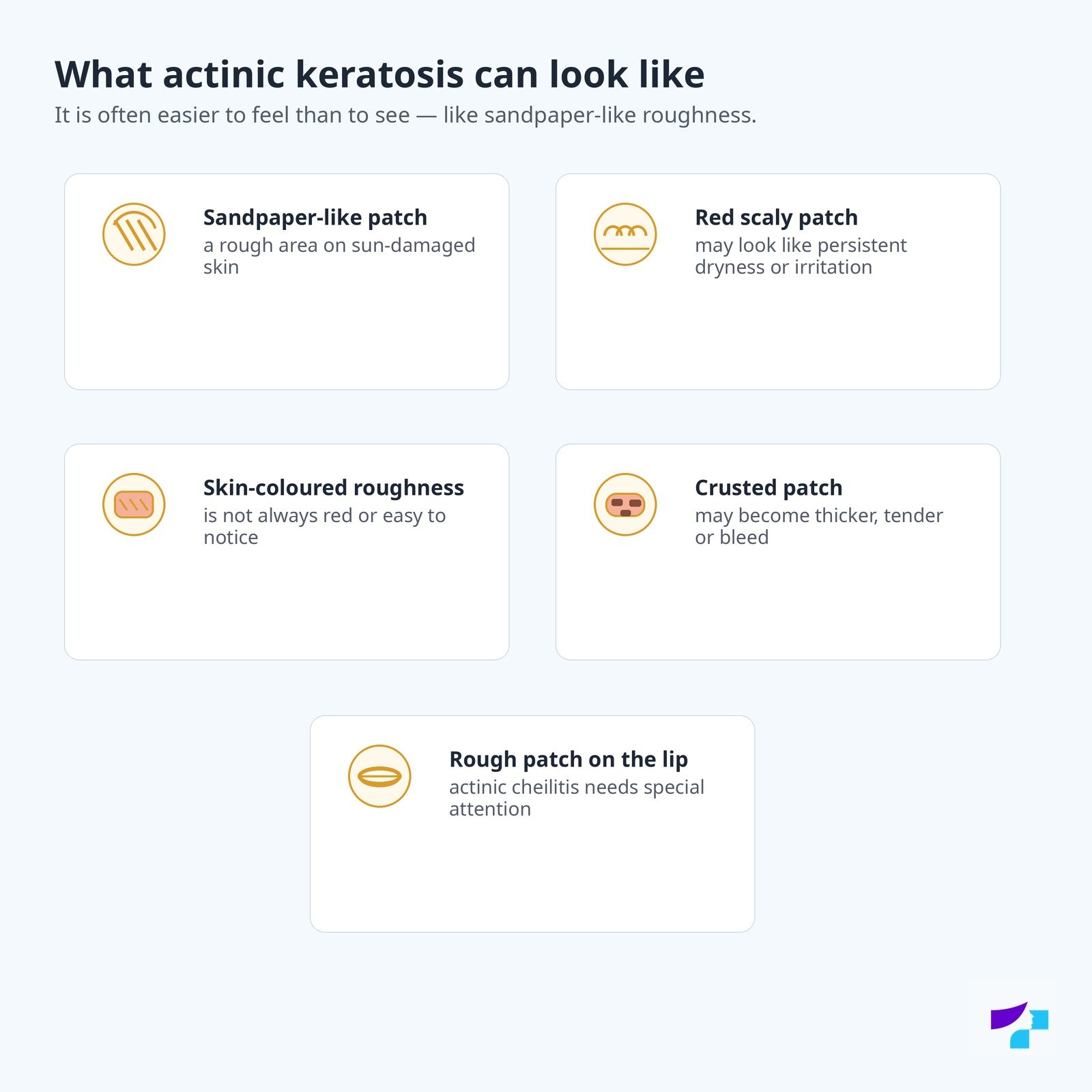
Actinic keratosis may appear as:
- a rough or sandpaper-like patch;
- a pink or red scaly patch;
- a skin-coloured rough area;
- brownish or pigmented roughness;
- a dry patch that does not go away;
- a crusted patch;
- tender or painful roughness;
- a rough patch on the lip;
- multiple rough patches in one sun-damaged area.
In the early stage, actinic keratosis can be very small. A person may notice only the sensation that the skin is rough in one spot, even though visually the patch seems almost unnoticeable. Some lesions may be visible, while others are only palpable. Therefore, during a dermatological examination, it is important not only to visually check the skin but also to assess the surface and location of the lesion.
Can actinic keratosis hurt, itch or bleed?
Actinic keratosis may cause no symptoms, but it may also itch, sting, hurt or become tender to touch. Sometimes it may become covered with a crust or bleed, especially after scratching, shaving or trauma.
Pain, bleeding, oozing, ulceration or rapid thickening are signs that the lesion should not be treated as simple dry skin. In such cases, a dermatologist’s examination is needed, because the lesion may have become suspicious for squamous cell carcinoma.
Three to five main signs that may suggest actinic keratosis
The following signs most often help recognise actinic keratosis.
Rough, sandpaper-like patch
The patch may be small, but it can be felt clearly with a finger as roughness on sun-damaged skin.
Scaling or dryness that does not go away
If an area of “dry skin” does not disappear with ordinary skin care, it should be assessed.
Pink, red or skin-coloured patch
Actinic keratosis is not always bright red. It may also be skin-coloured or slightly brownish.
Location on sun-damaged areas
Typical areas include the face, nose, forehead, ears, scalp, lips, backs of the hands and forearms.
Changes over time
If the patch becomes thicker, painful, bleeds, oozes, ulcerates or grows quickly, it should be shown to a dermatologist.
When can a rough patch on the skin be dangerous?
A rough patch may be dangerous if it changes or has signs that may suggest progression to squamous cell carcinoma. Particular attention should be paid to patches that:
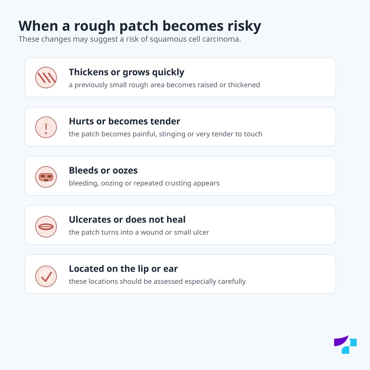
- become thicker;
- increase rapidly in size;
- hurt or become tender;
- bleed;
- ooze;
- form a repeated crust;
- ulcerate;
- do not improve after ordinary skin care;
- are located on the lip, ear, scalp, nose, backs of the hands or forearms;
- occur in a person with weakened immunity.
If such signs are present, it is not advisable to wait for a long time or treat the patch as ordinary dryness. If a rough patch becomes a raised nodule, ulcer, bleeding crust or painful thickening, it should no longer be considered ordinary actinic keratosis — squamous cell carcinoma must be excluded.
Where does actinic keratosis most often occur?
Actinic keratosis most often occurs in areas that have received a lot of sun exposure over a lifetime. Typical locations include:
- the face;
- the nose;
- the forehead;
- the cheeks;
- the ears;
- the lower lip;
- the scalp, especially in balding areas;
- the neck;
- the shoulders;
- the forearms;
- the backs of the hands.
Actinic keratosis can also occur in other areas, but it is less typical in places that are rarely exposed to the sun. If a rough or scaly patch appears in an unusual location, it should still be assessed based on its features rather than assuming that it is definitely not important.
Can actinic keratosis occur on the lip?
Yes. Actinic keratosis on the lip, especially on the lower lip, may appear as long-lasting dryness, roughness, cracking, crusting, tenderness or a whitish/pink area. This condition is often called actinic cheilitis.
Actinic cheilitis is sun damage in the lip area and may be associated with the risk of squamous cell carcinoma. Therefore, a long-lasting rough, cracked, bleeding or ulcerated lip should not be considered only “dryness”, “wind-chapped lips” or a cosmetic problem.
Particular caution is needed if the lower lip is persistently dry, scaly, cracked, loses its clear border with the surrounding skin, forms a crust or bleeds from time to time. In such cases, it is not advisable to use only lip balm or cosmetic products for a long time — an examination by a dermatologist or another appropriate specialist is needed.
Can actinic keratosis occur on the scalp?
Yes. Actinic keratosis can occur on the scalp, especially in people with thinning hair or baldness. This area is often exposed to the sun, but people check it less often themselves. Therefore, rough patches on the scalp are often noticed later.
A mirror can be used to examine the scalp, or help can be asked from a family member. If there are several rough patches, crusts or painful areas, a dermatologist should be consulted.
Why does actinic keratosis develop?
Actinic keratosis develops due to long-term exposure to ultraviolet radiation. UV radiation damages the DNA of skin cells. The body can repair some of this damage, but some damage accumulates over time. If the damage affects the mechanisms that control cell growth, the cells begin to behave abnormally and form a rough, scaly patch.
The main sources of risk are:
- the sun;
- tanning beds;
- long-term outdoor work;
- frequent outdoor sports or hobbies;
- repeated sunburns;
- insufficient sun protection over a lifetime.
Who has a higher risk of actinic keratosis?
Actinic keratosis can develop in anyone, but the risk is higher in certain groups.
Risk is increased in people who have:
- fair skin;
- blond or red hair;
- light-coloured eyes;
- skin that burns easily and tans poorly;
- long-term sun exposure over a lifetime;
- tanning bed use;
- outdoor work;
- frequent outdoor hobbies or sports;
- older age;
- many signs of sun damage on the skin;
- previous actinic keratosis;
- previous skin cancer;
- weakened immunity;
- organ transplantation or long-term immunosuppressive therapy.
Actinic keratosis is less common in people with darker skin, but it can still occur, especially with intense UV exposure, immunosuppression or other risk factors.
Can actinic keratosis be inherited?
Most actinic keratoses are not directly inherited. However, a person can inherit skin type, sensitivity to the sun and a tendency to burn easily. This may increase the risk that UV damage will accumulate more quickly over a lifetime. In rarer cases, genetic conditions that make the skin especially sensitive to UV damage may be associated with a higher risk of actinic keratosis and skin cancer.
Development mechanism of actinic keratosis
Actinic keratosis is not an infection, so it does not have a mechanism of infection. It is more accurate to speak about a development mechanism.
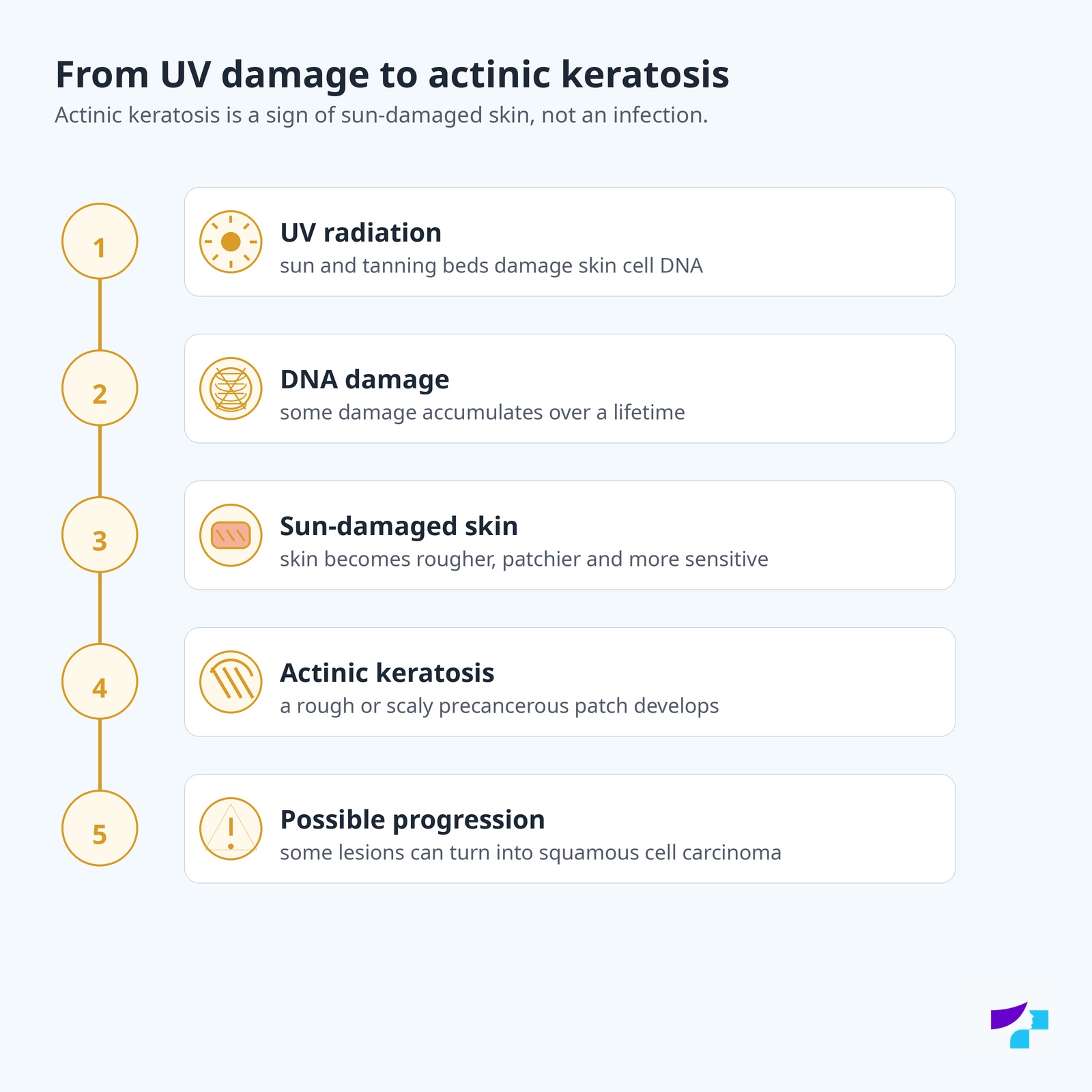
In simplified terms, it happens as follows:
- UV radiation from the sun or tanning beds damages the DNA of skin cells.
- Some damage is repaired, but some remains.
- Over time, damage accumulates in the sun-exposed field of skin.
- Keratinocytes become abnormal and begin to form a rough, scaly lesion.
- Actinic keratosis develops.
- In some cases, the lesion may progress to squamous cell carcinoma.
Timeline of actinic keratosis development
Actinic keratosis usually does not develop in one day. It is the result of long-term UV damage.
UV exposure
Years of exposure to the sun or tanning beds damage the DNA of skin cells.
Sun-damaged skin
The skin becomes rougher, more patchy, drier, with pigment changes, small blood vessels and signs of ageing.
Early roughness
A small sandpaper-like patch appears, which may be difficult to see.
Visible actinic keratosis
The patch becomes pink, red, skin-coloured, brownish, scaly or crusted.
Suspicious changes
If the lesion becomes thicker, painful, bleeds, oozes, ulcerates or grows quickly, squamous cell carcinoma must be excluded.
Actinic keratosis and skin cancer risk
Actinic keratosis is important not only as a single patch, but also as a signal that the skin is sun-damaged. The more actinic keratoses there are and the more extensive the sun damage, the higher the risk of cutaneous squamous cell carcinoma may be.
Not every actinic keratosis turns into cancer. However, it is not possible to reliably predict which lesion will progress. Therefore, a dermatologist assesses not only one patch but the overall condition of the skin: how many lesions there are, where they are located, whether they are changing, whether the patient is immunosuppressed and whether there has been previous skin cancer.
What is field cancerisation?
Field cancerisation means that long-term UV radiation has damaged not only one visible patch, but a wider area of skin. Actinic keratoses may be visible in this field, but some cellular changes may be microscopic and not yet visible.
For a patient, this can be explained simply: if there are many rough patches on the forehead, scalp, ears or backs of the hands, the problem is not limited to one patch. The whole area has been sun-damaged for a long time and may form new lesions in the future.
Some lesions may be visible as rough patches, while some cellular changes may not yet be visible to the eye. Therefore, a dermatologist may sometimes treat apparently normal skin around the visible patches if the whole area has been damaged by the sun for a long time.
That is why sometimes treatment is aimed not only at one patch, but at a wider area of sun-damaged skin. This is called field therapy. The doctor chooses the method according to the number of lesions, their location, the condition of the skin and the patient’s risk.
How can you tell that actinic keratosis is becoming dangerous?
More concerning signs may include:
- rapid growth;
- thickening;
- pain;
- bleeding;
- oozing;
- ulceration;
- recurrent crusting;
- marked tenderness;
- the lesion becomes nodule-like;
- the patch is located on the lip, ear or scalp, or occurs in the setting of immunosuppression.
Such changes may mean that the lesion is no longer just actinic keratosis and that squamous cell carcinoma must be excluded.
Can actinic keratosis become melanoma or basal cell carcinoma?
Actinic keratosis is mainly associated with the risk of squamous cell carcinoma, because both conditions arise from keratinocytes. It does not turn into melanoma, because melanoma arises from different cells — melanocytes. It is also not a typical precursor of basal cell carcinoma.
However, a person with actinic keratosis often has sun-damaged skin overall, and UV damage is also a risk factor for other skin cancers. Therefore, the dermatologist should assess the whole skin, not just one rough patch.
When should you see a dermatologist?
You should see a dermatologist if:
- a rough patch does not go away;
- the patch becomes thicker;
- the patch hurts, itches or becomes tender;
- the patch bleeds, oozes or ulcerates;
- a recurrent crust forms;
- the patch is located on the lip, ear, face, scalp or backs of the hands;
- there are many similar rough patches;
- the person has weakened immunity;
- there has been previous skin cancer;
- it is not clear whether the patch is actinic keratosis, squamous cell carcinoma, basal cell carcinoma or another skin disease.
How is actinic keratosis diagnosed?
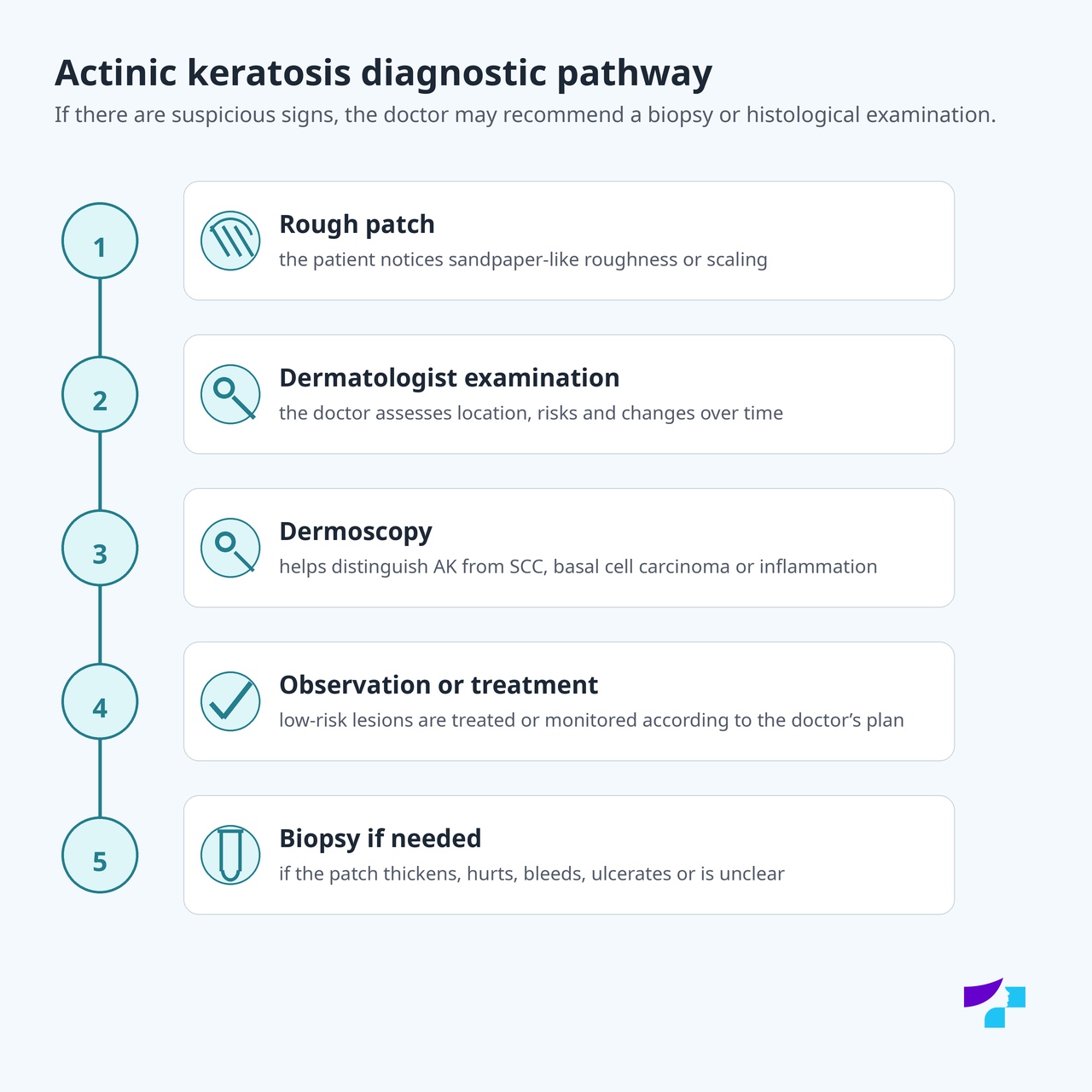
The dermatologist usually starts with a skin examination and assessment of the patient’s risk factors. The doctor evaluates the appearance, surface, roughness, colour, location, pain, bleeding, ulceration and changes over time.
Dermoscopy is often used. Dermoscopy is the examination of a skin lesion with a special optical device — a dermatoscope. It helps reveal structures that are not visible to the naked eye and distinguish actinic keratosis from other skin lesions.
Related topics are discussed in the articles Dermoscopy and Skin lesion examination.
When is a biopsy needed?
A biopsy means taking a tissue sample for microscopic examination. In actinic keratosis, biopsy is not always needed. It may be necessary if there are signs that raise suspicion of squamous cell carcinoma or another skin cancer.
A biopsy may be recommended if the patch:
- grows quickly;
- becomes thick or nodular;
- hurts;
- bleeds;
- ulcerates;
- oozes;
- repeatedly forms a crust;
- does not improve after treatment;
- is located in a high-risk area;
- looks atypical.
What is histological examination?
Histological examination is the microscopic examination of tissue. It helps determine whether the lesion is actinic keratosis, Bowen disease, squamous cell carcinoma, basal cell carcinoma or another diagnosis. Histology is important when it is not possible to reliably understand clinically how deep and serious the changes are.
Can actinic keratosis be diagnosed only from a photo?
No. A photograph may help show that a patch looks suspicious, but it does not replace a dermatologist’s examination, dermoscopy and, if needed, biopsy. Actinic keratosis can be confused with eczema, psoriasis, fungal infection, basal cell carcinoma, squamous cell carcinoma and other skin conditions.
What can actinic keratosis be confused with?
Actinic keratosis can be confused with:
- dry skin;
- eczema;
- psoriasis;
- fungal infection;
- seborrhoeic keratosis;
- a wart;
- basal cell carcinoma;
- squamous cell carcinoma;
- Bowen disease;
- a scar or chronic irritation;
- a pigmented skin lesion.
If the patch does not go away, grows, bleeds, hurts or becomes thicker, a dermatologist’s examination is needed.
How is actinic keratosis treated?
Treatment of actinic keratosis depends on the number, size, thickness and location of the lesions, the patient’s risk, and whether one patch or a wider field of sun-damaged skin needs to be treated.
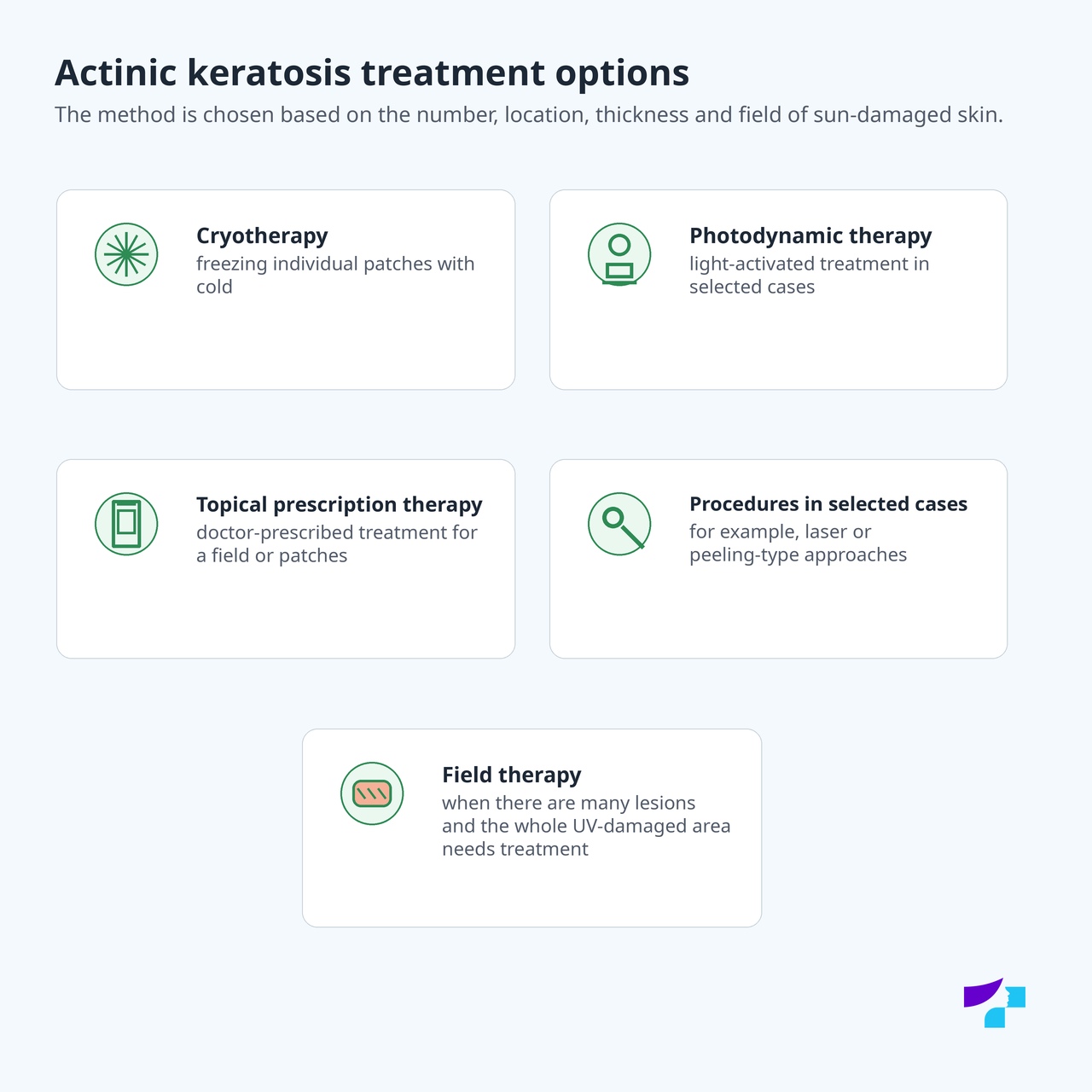
The main treatment options are:
- cryotherapy, or freezing;
- photodynamic therapy;
- topical prescription therapy prescribed by a doctor;
- curettage or surgical removal in selected cases;
- laser or chemical peel-type procedures in selected situations;
- field therapy if there are many lesions or the whole area is sun-damaged;
- regular monitoring in selected low-risk cases after medical assessment.
A moisturising cream or cosmetic skin care may temporarily reduce dryness and discomfort, but it does not treat the abnormal cellular changes if the patch is actinic keratosis. If the roughness does not go away or recurs in the same place, a dermatologist’s assessment is needed.
This article does not list specific prescription medicine names. Specific treatment is determined by a doctor after assessing the skin condition, lesion risk and the patient’s individual situation.
Does actinic keratosis need to be treated?
Actinic keratosis is often recommended to be treated because it can progress and indicates a field of sun-damaged skin. The aims of treatment are to:
- remove the specific lesion;
- reduce the risk of progression;
- reduce the formation of new lesions in the sun-damaged area;
- improve skin monitoring;
- notice dangerous changes in time.
However, not every lesion is treated in the same way. In some cases, the doctor may recommend monitoring, in others treatment of a single patch, and in others field therapy for the whole area.
When is one patch treated, and when is the whole sun-damaged area treated?
One patch is more often treated when actinic keratosis is isolated, clearly visible and there are not many similar lesions.
A wider area is treated when there are many actinic keratoses or a visible field of sun-damaged skin. In this case, the problem is not only one patch — abnormal cellular changes may also be present in the surrounding skin. Therefore, the doctor may recommend treatment that acts on the whole area.
Is actinic keratosis treated with cryotherapy?
Yes. Cryotherapy, or freezing, is one of the most commonly used methods for treating individual actinic keratoses. After the procedure, the skin may become red and tender, form a blister or crust, and then heal. Cryotherapy is not suitable for all lesions, and its suitability is determined by the doctor.
Is actinic keratosis treated with photodynamic therapy?
Yes. Photodynamic therapy may be suitable for certain forms of actinic keratosis and for a wider field of sun-damaged skin. This approach uses a special substance and light to damage abnormal cells. After treatment, the skin may be red, tender and peel. The method is chosen by the doctor.
Is actinic keratosis treated with cream?
Yes, in certain cases actinic keratosis is treated with topical prescription therapy prescribed by a doctor. This type of therapy can be especially useful when there are many lesions or when a wider field of sun-damaged skin needs treatment. During treatment, the skin may become red, inflamed, tender or peel — this can be an expected treatment effect, but treatment must be carried out under medical guidance.
Can actinic keratosis be treated with laser or chemical peeling?
In selected cases, a dermatologist may consider laser or chemical peel-type procedures, especially for treating wider sun-damaged skin. They are not suitable for all patients and all lesions. If invasive cancer is suspected, accurate diagnosis is needed first, not a cosmetic procedure.
Will there be a scar after treatment?
After some treatment methods, temporary redness, crusting, peeling, pigment changes or tenderness may occur. The risk of scarring usually depends on the method, lesion depth, location, skin characteristics and healing. If the lesion needs to be excised or a deeper procedure is needed, the risk of scarring may be higher.
How long does the skin take to heal after actinic keratosis treatment?
Healing time depends on the treatment method. After cryotherapy, the skin may heal within a few weeks. After topical field therapy, redness, tenderness and peeling may last longer. After photodynamic therapy or procedures, healing time is individual. Precise care instructions are provided by the treating doctor.
Can actinic keratosis be treated at home?
Actinic keratosis should not be treated at home with self-selected remedies, acids, burning, scratching or preparations recommended online. Self-treatment may delay diagnosis, especially if the lesion is in fact already squamous cell carcinoma or another skin cancer.
At home, it is useful and necessary to follow sun protection, check the skin and follow the doctor’s instructions, but the treatment method should be determined by a dermatologist.
What should not be done if a patch may be actinic keratosis?
A suspicious rough or scaly patch should not be:
- burned with acids or folk remedies;
- scraped off or traumatised;
- treated for a long time as dry skin if there is no improvement;
- removed by a cosmetic procedure without a diagnosis;
- treated with prescription medicines without a doctor’s instruction;
- assessed only by internet pictures;
- ignored if the patch hurts, bleeds, oozes, ulcerates or grows quickly.
What happens if actinic keratosis is not treated?
If actinic keratosis is not treated, it may persist, improve or progress. Some patients may develop new lesions in the same sun-damaged area. Some lesions may turn into squamous cell carcinoma.
Untreated actinic keratosis does not always mean that cancer will definitely develop, but it increases the need for dermatological assessment and follow-up.
Can you die from actinic keratosis?
Actinic keratosis itself is not fatal. However, its significance is that it may be a precancerous lesion and indicates sun-damaged skin. If the lesion progresses to invasive squamous cell carcinoma and is not treated, the risk may become much more serious.
What is the prognosis for actinic keratosis?
The prognosis is usually good if lesions are recognised and treated in time and the skin is protected from further UV damage. However, actinic keratosis may recur, and a person with many actinic keratoses has a higher risk of skin cancer. Therefore, treatment of a single patch is not enough — prevention and follow-up are also important.
How can the risk of actinic keratosis be reduced?
The risk of actinic keratosis can be reduced by decreasing the impact of UV radiation on the skin.
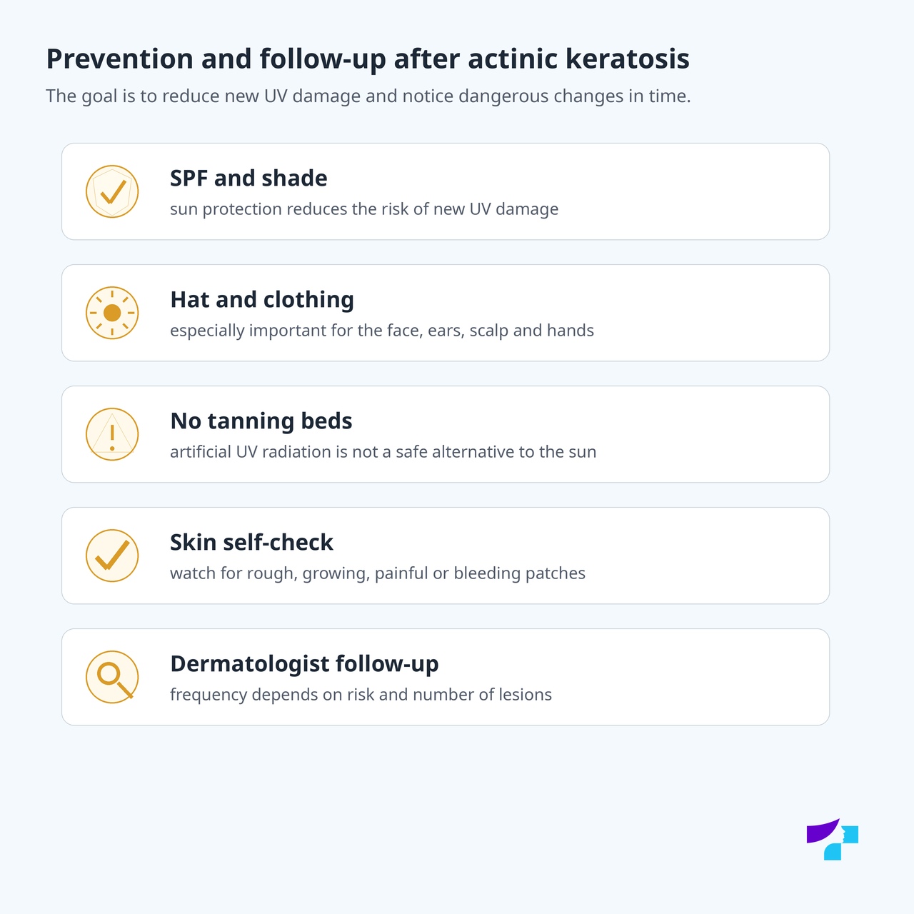
The most important prevention principles are:
- avoid sunburn;
- do not use tanning beds;
- seek shade during intense UV radiation;
- wear a wide-brimmed hat;
- wear sunglasses;
- choose clothing that covers the skin;
- use a broad-spectrum sunscreen with appropriate SPF;
- reapply sunscreen after swimming, sweating or towel-drying;
- especially protect the face, ears, lips, scalp, neck and backs of the hands;
- check the skin regularly;
- visit a dermatologist if there is extensive sun damage or previous skin cancer.
The safest approach is a combination of several layers of protection: avoiding intense sun, shade, skin-covering clothing, a hat, sunglasses, SPF products and not using tanning beds. SPF alone is not sufficient if a person deliberately extends time spent in the sun.
Does SPF help reduce the risk of actinic keratosis?
Yes, properly used SPF helps reduce skin damage caused by UV radiation. However, SPF is not the only protection and is not permission to stay in the sun longer. Combined protection works best: shade, clothing, a hat, sunglasses, SPF and avoiding tanning beds.
Can you sunbathe after a diagnosis of actinic keratosis?
Intentional sunbathing is not recommended. Actinic keratosis means that the skin is already sun-damaged. This does not mean that a person cannot go outside, but sunburn, tanning beds and prolonged intense UV exposure should be avoided.
Should children use sun protection?
Yes. Children’s skin should be protected from sunburn because UV damage accumulates over a lifetime. Actinic keratosis more often appears later in life, but its basis is often UV damage accumulated over many years. Children should use shade, clothing, hats and age-appropriate sun protection products.
How to check your skin at home?
The skin should be checked in good lighting, using a mirror or asking a family member for help.
Particular attention should be paid to:
- the face;
- the nose and forehead;
- the ears;
- the lips;
- the scalp;
- the neck;
- the shoulders;
- the forearms;
- the backs of the hands;
- areas where rough patches or treated lesions have previously been present.
Attention should be paid to patches that are rough, scaly, thickening, bleeding, painful, oozing, ulcerated or persistent.
How often should the skin be checked by a dermatologist?
The frequency of check-ups is determined by the dermatologist. It depends on the number of lesions, the patient’s skin type, the extent of UV damage, immune system status, previous skin cancer and treatment results. People with many actinic keratoses, immunosuppression or previous skin cancer usually need more frequent follow-up.
What is well established in medicine at present?
It is medically well established that actinic keratosis is associated with cumulative UV radiation damage. It is also well established that actinic keratosis is associated with the risk of squamous cell carcinoma, especially in people with many actinic keratoses, extensive sun damage, previous skin cancer or weakened immunity.
An evidence-based approach includes dermatologist examination, dermoscopy, biopsy in suspicious cases, treatment of a single lesion or field therapy, as well as long-term UV protection and follow-up.
What is still being discussed and developed in medicine?
Medicine continues to improve risk assessment: which actinic keratosis lesions are the most dangerous, which patients need more intensive treatment and how best to reduce the risk of squamous cell carcinoma. Digital dermoscopy, skin photography, artificial intelligence support tools and personalised risk assessment are developing.
Field therapy approaches are also being studied and refined. These treat not only individual patches but the entire sun-damaged skin field. The aim is to reduce the recurrence of new actinic keratoses and possibly reduce the risk of squamous cell carcinoma.
References and authoritative sources
- Centre for Disease Prevention and Control of Latvia (SPKC): Don’t burn! Choose to live!
- Centre for Disease Prevention and Control of Latvia (SPKC): Safe sun exposure
- UK National Health Service (NHS): Actinic keratoses / solar keratoses
- British Association of Dermatologists: patient information on actinic keratoses
- DermNet: Actinic keratosis
- DermNet: Cutaneous squamous cell carcinoma
- US National Cancer Institute (NCI): skin cancer treatment — patient information
- American Academy of Dermatology Association (AAD): actinic keratosis — overview and treatment
- Cancer Research UK: risks and causes of non-melanoma skin cancer, including solar keratosis
- PubMed Central: actinic keratosis and cutaneous squamous cell carcinoma
- PubMed: field cancerisation therapies in the management of actinic keratosis
Disclaimer
The information in this article is intended for informational and educational purposes and does not replace medical consultation, diagnosis or treatment. Self-assessment of skin patches and lesions can be wrong, because actinic keratosis may resemble dry skin, eczema, psoriasis, fungal infection, basal cell carcinoma, squamous cell carcinoma or other skin changes. If you notice a new, rough, scaly, growing, bleeding, painful, itchy, oozing, ulcerated, crusted, non-healing or otherwise suspicious skin or lip patch, consult a dermatologist, dermato-oncologist or another appropriate specialist. Do not try to burn, scratch, cut, scrape off or treat suspicious patches with self-selected remedies. If the lesion grows rapidly, bleeds significantly, ulcerates, becomes very painful, shows signs of infection, enlarged lymph nodes or general symptoms appear, urgent medical assessment is needed.
