A skin lesion examination is an assessment of the skin and individual skin lesions performed by a dermatologist. During the examination, the doctor inspects the skin, assesses specific lesions and, when needed, uses dermoscopy — examination of a skin lesion under magnification and special lighting.
A skin lesion examination is not limited to a mole check. The dermatologist also assesses warts, papillomas, keratomas, seborrhoeic keratoses, angiomas, fibromas, pigmented patches, pink or skin-coloured nodules, non-healing crusts, sores and other lesions whose nature is unclear to the patient.
The purpose of the examination is to determine whether a lesion appears benign, whether it should be monitored, removed, examined histologically or referred for further treatment. It is particularly important to examine lesions that grow, change colour or shape, bleed, itch, hurt, ulcerate or look different from other lesions on the skin.
Why is a skin lesion examination important?
Many skin lesions are benign and pose no medical threat. However, benign, precancerous and malignant skin lesions can sometimes look similar. At home, patients often cannot reliably distinguish a mole from a seborrhoeic keratosis, a papilloma from a wart or a benign pigmented lesion from an early melanoma.
A skin lesion examination is important because:
- some skin cancers cause no pain or discomfort at an early stage;
- suspicious changes may be visible on the skin but require professional assessment;
- dermoscopy allows the dermatologist to see structures that cannot be assessed reliably with the naked eye;
- digital dermoscopy helps store images and compare changes in lesions over time;
- before a skin lesion is removed, it is essential to determine whether cosmetic removal is appropriate or histological examination is required.
Skin cancer is not one specific type of lesion. The most common types include basal cell carcinoma, squamous cell carcinoma and melanoma. Melanoma is less common than basal cell carcinoma and squamous cell carcinoma, but it has a greater risk of spreading, or metastasising. Timely diagnosis of suspicious skin lesions is therefore especially important.
Which skin lesions does a dermatologist examine?
A dermatologist examines more than moles. During a skin lesion examination, the doctor may assess lesions on the face, body, scalp, neck, back, arms, legs, feet, palms and other areas of the skin.
A dermatologist can examine:
- moles — pigmented lesions that are present from birth or acquired during life;
- pigmented lesions — darker patches, nodules or unevenly coloured lesions;
- non-pigmented lesions — skin-coloured, pale, pink, scaly or nodular lesions;
- warts — skin growths that are often caused by a virus;
- papillomas — soft, skin-coloured growths that commonly occur on the neck, in the armpits or in skin folds;
- keratomas and seborrhoeic keratoses — often benign, rough, brownish or greyish lesions;
- angiomas — red vascular lesions;
- fibromas — benign connective-tissue lesions;
- non-healing crusts, ulcers or bleeding lesions;
- suspicious new skin growths whose origin is unclear to the patient.
Some benign lesions may look alarming, while some malignant lesions may initially appear inconspicuous. The main task is therefore not to name the lesion yourself, but to notice whether it is new, changing, different or symptomatic.
Who is particularly advised to have skin lesions examined?
A skin lesion examination is advisable for anyone who notices a new, changing or unclear lesion. It is especially important for people with an increased risk of skin cancer.
Higher-risk groups include people who have:
- fair skin that burns easily in the sun;
- fair or red hair, light-coloured eyes or many freckles;
- many moles;
- atypical or irregular moles;
- frequent sunburn, particularly during childhood or adolescence;
- long-term or intense exposure to ultraviolet radiation;
- a history of tanning-bed use;
- a personal or family history of skin cancer;
- a weakened immune system;
- long-term outdoor work in the sun;
- chronic non-healing skin damage;
- previously treated precancerous changes or skin tumours.
Ultraviolet radiation is one of the most important preventable risk factors for skin tumours. The risk increases with exposure both to sunlight and to artificial ultraviolet radiation, including tanning beds.
When should you not wait to have a skin lesion checked?
You should see a dermatologist promptly if a lesion:
- grows rapidly;
- changes colour, shape or borders;
- becomes asymmetrical;
- bleeds without a clear injury;
- forms an ulcer or a non-healing crust;
- itches, hurts, stings or becomes tender;
- appears in adulthood and continues to change;
- looks markedly different from other lesions on the skin;
- is repeatedly injured by clothing, shaving, a comb or jewellery;
- simply causes uncertainty or concern.
Pain is not a reliable criterion. A suspicious skin lesion may initially be painless, not itch and cause no discomfort. You should therefore not wait until pain, bleeding or marked inflammation develops.
Five signs that a lesion should be checked
It is advisable to book an appointment with a dermatologist if any of the following signs are present:
- The lesion is changing — it becomes larger, darker, lighter, redder or more uneven.
- Its shape or borders change — it becomes asymmetrical or develops blurred or notched edges.
- The lesion bleeds or ulcerates — especially when the bleeding is not associated with a clear injury.
- A non-healing crust forms — the crust falls off and then forms again.
- The lesion differs from the others — it looks unlike the other moles or skin lesions.
These signs do not automatically mean cancer, but they are sufficient reason for a professional examination.
The ABCDE rule for self-examination of pigmented lesions
The ABCDE rule is a simple way to notice changes in pigmented lesions. It does not replace a dermatologist’s examination, but it helps patients understand when a lesion should be shown to a doctor.
| Letter | Feature | What should you look for? |
|---|---|---|
| A | Asymmetry | One half of the lesion differs significantly from the other. |
| B | Border | The borders are notched, blurred, irregular or poorly defined. |
| C | Colour | Several colours are visible in the lesion or its colour becomes uneven. |
| D | Diameter | The lesion is growing or is larger than before. Change over time is more important than any single size threshold. |
| E | Evolving | The lesion changes in size, shape, colour or surface, or begins to cause symptoms. |
The ABCDE rule is particularly useful for self-examination of moles and other pigmented lesions, but it is not a complete diagnostic method. Some dangerous lesions may not be typically dark or classically pigmented.
What is the “ugly duckling” sign?
The “ugly duckling” sign is also important during skin self-examination. It means paying attention to a lesion that looks different from the other lesions on that particular person’s skin.
People usually have a recognisable pattern of moles and pigmentation, with similarities in colour, shape, size or distribution. A lesion that differs markedly from this pattern should be shown to a dermatologist.
This principle is particularly useful for people with many moles. The suspicious lesion may not be the largest or darkest one, but the one that looks different from the others.
Does skin cancer always look like a dark mole?
No. Melanoma may be dark and unevenly pigmented, but some malignant skin lesions may be pink, pale, skin-coloured, scaly or appear as a nodule or non-healing wound.
Basal cell carcinoma may look like a shiny nodule, a pink patch, a non-healing crust or a small ulcer. Squamous cell carcinoma may appear as a rough, scaly, thickened or bleeding lesion.
Attention should therefore be paid not only to dark moles, but also to any new, growing, bleeding, ulcerated or non-healing skin lesion.
Are all suspicious skin lesions skin cancer?
No. Many lesions that look suspicious turn out to be benign. For example, a seborrhoeic keratosis may be dark, rough and irregular but is often benign. Warts, papillomas, angiomas and fibromas are also usually benign.
However, the nature of a lesion cannot always be determined reliably by looking at it with the naked eye. The purpose of a skin lesion examination is not to create fear, but to distinguish situations in which observation is sufficient from those requiring dermoscopy, removal or histological examination.
How is a skin lesion examination performed?
A skin lesion examination usually takes place in several stages.
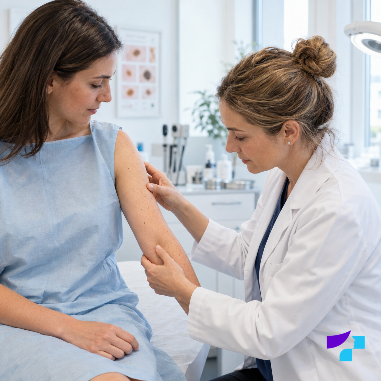
During a clinical examination, the dermatologist assesses the appearance of skin lesions and the surrounding skin.
Discussion with the doctor
At the beginning of the appointment, the dermatologist asks which lesion concerns the patient, how long it has been present and whether it has changed, bled, itched, hurt or been injured.
The doctor may also ask about:
- sunburn;
- tanning-bed use;
- skin cancer in the family;
- previously removed lesions;
- previous dermoscopy examinations;
- diseases or treatments that affect the immune system;
- changes in skin lesions during recent months.
Skin examination
The doctor examines the specific lesion and, when necessary, a wider area of skin or several lesions. A patient may attend because of one concern, but the dermatologist may recommend assessing other lesions as well, particularly when the patient has many moles or an increased risk of skin cancer.
Dermoscopy
If the nature of a lesion cannot be assessed reliably by ordinary inspection, or if it is pigmented, changing, atypical, pink, scaly or non-healing, the doctor may use a dermatoscope.
A dermatoscope helps reveal the lesion’s colour, pigment distribution, borders, vascular pattern and other structures that are not clearly visible to the naked eye.
The doctor’s conclusion and further plan
After the examination, the dermatologist explains whether the lesion appears benign and whether it should be monitored, removed or sent for histological examination.
In simplified terms, the process is: dermatologist examination → dermoscopy when needed → a plan for monitoring, removal or additional investigation.
A skin lesion can be assessed during a dermatologist consultation.
Are the results available immediately?
The dermatologist usually explains the initial conclusion during the appointment. The doctor can say whether the lesion appears benign and whether it should be monitored, removed or investigated further.
However, two things must be distinguished:
- the dermatologist’s visual and dermoscopic assessment, which can usually be explained during the appointment;
- the final tissue diagnosis, which in suspicious cases is confirmed by histological examination after removal of the lesion.
If histology is needed, the final result will be available after laboratory examination.
Is a skin lesion examination the same as dermoscopy?
No. A skin lesion examination is a broader process, while dermoscopy is one method that a dermatologist may use during that process.
| Skin lesion examination | Dermoscopy |
|---|---|
| Includes assessment of the patient’s concerns and risk factors. | Is a specific non-invasive examination method. |
| Includes clinical inspection of the skin and lesion. | The lesion is examined under magnification with an optical device and special lighting. |
| May include dermoscopy when needed. | Helps assess pigment, blood vessels and other structures that are difficult to see with the naked eye. |
| Ends with a decision about monitoring, removal, histology or further treatment. | Does not always provide the final tissue diagnosis on its own. |
What is dermoscopy?
Dermoscopy is a non-invasive method for diagnosing skin lesions. Non-invasive means that the skin is not cut and no tissue sample is taken during the procedure. The dermatologist uses a dermatoscope to examine the lesion under magnification and special lighting.
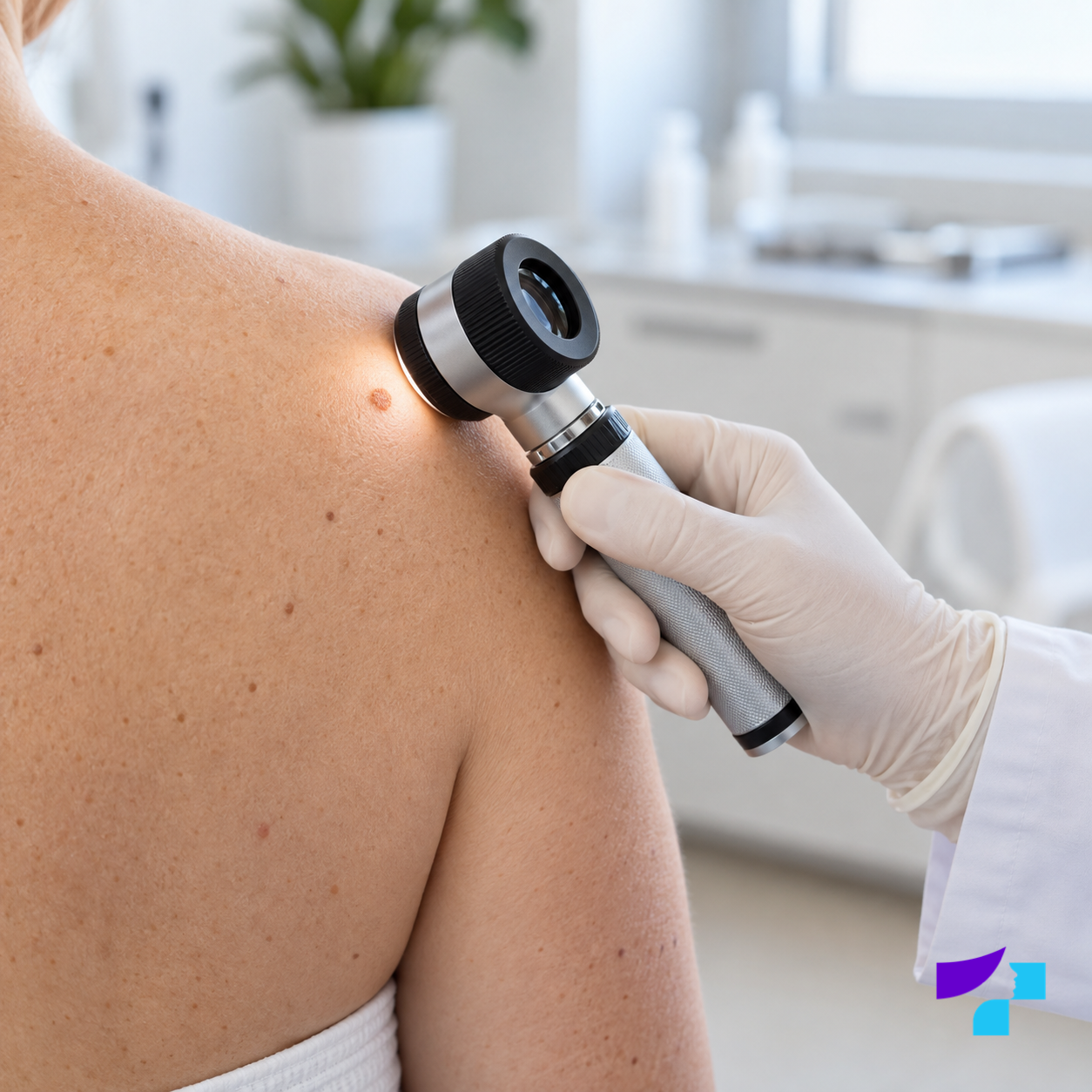
Dermoscopy allows the doctor to assess the structure of a skin lesion and features that cannot be seen with the naked eye.
Dermoscopy helps assess:
- pigment distribution;
- variation in colour;
- the lesion’s borders;
- the vascular pattern;
- surface structures;
- features that may suggest a benign, precancerous or malignant process.
Dermoscopy is especially important when assessing pigmented lesions, moles and melanoma risk, but it is also used for various non-pigmented, pink, scaly or vascular lesions.
Optical and digital dermoscopy
Optical dermoscopy
Optical dermoscopy is the direct examination of a lesion using a handheld dermatoscope. The doctor sees the lesion under magnification and assesses its structure during the appointment. This method is suitable for evaluating one or several specific skin lesions.
Digital dermoscopy
Digital dermoscopy makes it possible to capture an image of the lesion, enlarge it on a screen, store it and compare it with images taken at later appointments.
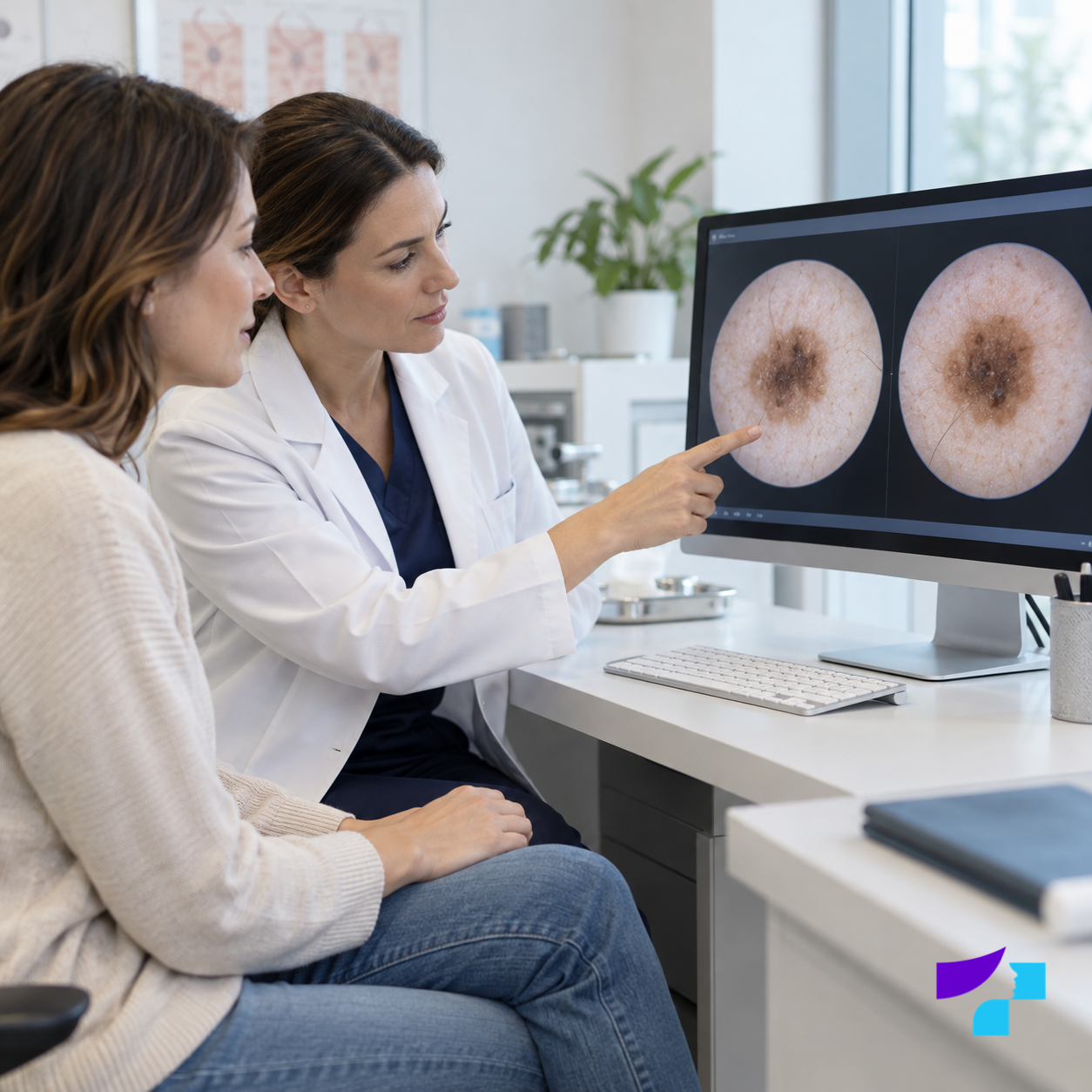
Digitally stored images help compare a skin lesion at different appointments and identify changes over time.
Digital dermoscopy is particularly useful when:
- the patient has many moles;
- there are atypical or difficult-to-assess lesions;
- lesions need to be monitored over time;
- small changes need to be followed;
- the patient has an increased risk of melanoma or another skin cancer;
- suspicious or malignant skin lesions have previously been removed.
Digital dermoscopy does not replace the doctor’s clinical judgement. It is a tool that helps document and compare lesions.
What is skin lesion mapping?
Skin lesion mapping is the systematic documentation and comparison of skin lesions over time. It may include photography of the whole body or selected areas, digital dermoscopy and comparison of images at subsequent appointments.
Skin lesion mapping is particularly useful for patients who have:
- a very large number of moles;
- atypical moles;
- a personal history of skin cancer;
- a family history of melanoma;
- changes that are difficult to notice during self-examination;
- a need to monitor several lesions at the same time;
- an increased risk of skin cancer.
Digital skin imaging systems help the doctor store clinical and dermoscopic images, compare changes over time and identify lesions that require closer monitoring.
Photographs taken at home may help a patient notice changes, but they do not replace medical dermoscopy. Lighting, distance, camera quality and angle can significantly affect an image.
What is histological examination?
Histological examination is the microscopic assessment of a removed tissue sample. It is performed in a laboratory to determine the precise nature of the lesion and check whether malignant cells are present.
Dermoscopy helps the doctor assess a lesion, but suspicious cases may require histology for a final diagnosis.
Histology is particularly important when:
- the lesion is suspicious;
- it changes rapidly;
- it bleeds or ulcerates;
- the dermatologist cannot reliably rule out a malignant process;
- the lesion is removed for medical reasons;
- a dangerous process must be excluded before cosmetic removal.
Why should a skin lesion be checked before removal?
Before removing a skin lesion, a dermatologist must assess whether it is benign and which removal method is safest. Not every lesion is suitable for laser treatment, electrocoagulation, cryotherapy or another cosmetic method.
An examination before removal helps determine:
- whether the lesion is benign;
- whether it may be removed with a laser or another cosmetic method;
- whether surgical removal is needed;
- whether histology should be performed after removal;
- whether similar lesions elsewhere on the body should also be checked.
If a lesion is pigmented, changing rapidly, bleeding or otherwise suspicious, surgical removal with histological examination may be the safer option. A suspicious lesion must not be destroyed solely for cosmetic reasons, as this may remove the opportunity to examine the tissue accurately in a laboratory.
Read more about the treatment of specific benign lesions in the articles on mole removal, wart removal and papilloma removal.
Can a skin lesion be removed with a laser?
A skin lesion should be removed with a laser only after a dermatologist has assessed it and confirmed that the specific lesion is suitable for this method. Laser treatment may be appropriate for selected benign lesions, but not for every lesion.
A suspicious, pigmented, rapidly changing or unclear lesion must not simply be “burned off”, treated with caustic substances, tied off or removed cosmetically without diagnosis. This may eliminate the possibility of complete histological examination and leave a dangerous process insufficiently assessed.
If the lesion is suspicious, the doctor may recommend surgical removal with histological examination rather than a laser procedure.
What happens if a skin lesion appears suspicious?
A suspicious finding does not mean that the patient definitely has cancer. It means that the lesion requires closer assessment or a safer next step.
The dermatologist may recommend:
- a repeat examination after a specified period;
- digital monitoring and comparison of images;
- surgical removal of the lesion;
- histological examination;
- referral to a surgeon, oncologist or another specialist;
- additional investigations when needed.
It is important to follow the doctor’s recommendations and continue monitoring when a lesion has been identified as requiring follow-up over time.
How often should skin lesions be checked?
There is no single examination interval suitable for everyone. The frequency is determined individually, taking into account skin type, the number of moles, previous lesions, family history, ultraviolet exposure and other factors.
For a person without increased risk, regular self-examination and a dermatologist appointment when a new or changing lesion appears are usually most important.
For people at increased risk, the dermatologist may recommend more regular checks, such as periodic dermoscopy or digital monitoring of lesions.
Skin self-examination at home
Skin self-examination helps detect changes early, but it does not replace a dermatologist. It is best performed in good lighting, using a mirror or asking someone close to help examine the back, scalp and other areas that are difficult to see.
During self-examination, pay attention to:
- new lesions;
- lesions that are changing;
- moles with ABCDE features;
- lesions that differ from the others;
- non-healing crusts or ulcers;
- bleeding or repeatedly injured lesions.
Self-examination should cover not only the face and arms, but also the back, shoulders, chest, abdomen, legs, feet, palms, spaces between the fingers and toes, scalp and skin folds.
For patients with many moles, taking photographs under consistent conditions may be useful, but photographs taken at home are not a complete diagnostic method.
Skin lesion examination in children
Skin lesions in children may change as they grow. Not every change automatically indicates a dangerous process. However, a child should be shown to a dermatologist if a lesion:
- grows rapidly;
- bleeds;
- hurts or itches;
- looks very atypical;
- is repeatedly injured;
- causes concern for the parents;
- appears suddenly and continues to change.
Skin lesions in children should not be treated at home with caustic substances or removed without medical assessment.
Skin lesion examination during pregnancy
Hormonal changes during pregnancy may affect skin pigmentation and some lesions. Pregnancy is not, however, a reason to postpone the examination of a suspicious lesion.
Consult a dermatologist if a lesion:
- increases rapidly in size;
- becomes uneven;
- bleeds;
- ulcerates;
- changes colour;
- becomes painful;
- looks different from the other lesions.
The dermatologist will determine which approach is safe and necessary in the individual situation.
Skin lesions after sun exposure and tanning-bed use
Ultraviolet radiation from the sun and tanning beds damages skin cells and increases the risk of skin tumours. After intense sun exposure or sunburn, it is advisable to pay attention to whether any skin lesions have changed.
It is particularly important to check lesions that have become darker, irregular, tender, bleeding or rapidly growing after the summer.
A tanning bed is not a safe alternative to sunlight. Artificial ultraviolet radiation is a significant risk factor for skin damage and skin cancer.
Important ways to protect the skin include:
- avoiding sunburn;
- seeking shade during the hours of strongest sunlight;
- wearing protective clothing, a hat and sunglasses;
- using a suitable sunscreen;
- not using tanning beds;
- checking the skin regularly, especially after the summer season.
How should you prepare for a skin lesion examination?
No special preparation is usually required, but a few simple steps can help make the appointment more thorough.
Before the appointment, it is advisable to:
- avoid applying make-up to the area being examined;
- avoid self-tanning products immediately before the examination;
- consider which lesions have changed;
- remember when the changes were first noticed;
- bring previous dermoscopy images, medical reports or histology results, if available;
- prepare questions for the doctor;
- wear comfortable clothing if a broader skin examination is needed.
Do not scratch, burn, treat with caustic substances or attempt to remove a lesion before the appointment, as this may make assessment more difficult.
How much does a skin lesion examination cost?
The cost of a skin lesion examination may vary. It can be affected by:
- the doctor’s qualifications;
- the type of appointment;
- the number of lesions examined;
- the extent of dermoscopy required;
- whether one lesion or several are being examined;
- whether digital documentation is required;
- whether further treatment or lesion removal is needed.
It is therefore more accurate to describe the cost as the combined price of the appointment and any necessary procedures rather than one universal amount. When several lesions must be examined, the cost and duration of the appointment may differ from an assessment of one specific lesion.
What is well established in medicine, and where is technology developing?
What is well established
It is well established that ultraviolet radiation damages the skin and increases the risk of skin tumours. The value of dermoscopy in dermatological assessment of skin lesions is also established, particularly for pigmented lesions and melanoma risk.
Individual medical assessment
Individual assessment remains essential when a lesion is not straightforward. The decision between monitoring, repeat dermoscopy, removal or histology depends on the lesion’s appearance, the patient’s risk factors and changes over time.
Developing technologies
Digital dermoscopy, skin lesion mapping, image comparison over time and computer-assisted image analysis are developing in modern dermatology. These technologies can help doctors structure monitoring, but they do not replace clinical assessment by a dermatologist.
Medical disclaimer
This information is provided for informational purposes and does not replace a consultation with a dermatologist, diagnosis or treatment. If you notice a new, changing, bleeding, painful, ulcerated, itchy or otherwise suspicious skin lesion, consult a dermatologist. Do not attempt to cut, burn, injure, cauterise or treat a suspicious skin lesion at home.
References and authoritative sources
- Pauls Stradiņš Clinical University Hospital: examination of skin lesions using digital dermoscopy
- Health Center 4: self-examination of skin lesions
- ARS Medical Clinic: dermoscopy
- Dr. Mauriņš Clinic: skin diagnostics
- Azaryan Medical Clinic: diagnosis of skin lesions
- Derma Clinic Riga: skin lesion mapping using the FotoFinder system
- Latvian Centre for Disease Prevention and Control: correct use of sunscreen and skin cancer prevention
- World Health Organization (WHO): ultraviolet radiation and skin cancer risk
- International Agency for Research on Cancer (IARC): ultraviolet-emitting tanning devices as a carcinogenic factor
- U.S. National Cancer Institute: skin cancer types, risk factors and treatment principles
- American Academy of Dermatology (AAD): the ABCDEs of melanoma detection
- DermNet: skin biopsy and histological examination
- Jensen and Elewski: the ABCDE rule and the ugly duckling sign in melanoma detection
