Squamous cell carcinoma, or cutaneous squamous cell carcinoma, is a malignant skin tumour that arises from the flat skin cells called keratinocytes. These cells form the upper layer of the skin and help protect the body from the external environment. When DNA damage accumulates in these cells and they begin to grow uncontrollably, squamous cell carcinoma can develop.
Squamous cell carcinoma belongs to the group of non-melanoma skin cancers. The most common non-melanoma skin cancers are basal cell carcinoma and squamous cell carcinoma. Squamous cell carcinoma is not the same as basal cell carcinoma, and it is not the same as melanoma. It is usually more dangerous than basal cell carcinoma because it has a higher risk of spreading, or metastasising, especially when the tumour is large, deep, recurrent, located on the lip, ear, scalp, fingers, feet, in a scar or chronic wound, or when the patient has a weakened immune system.
Squamous cell carcinoma most often develops on sun-damaged skin: the face, ears, lower lip, scalp, neck, backs of the hands, forearms and other areas that have received a lot of ultraviolet radiation over a lifetime. However, it can also develop in chronic wounds, scars, areas of inflammation or long-term irritation, as well as on mucous membranes, for example in the mouth or genital area.
Squamous cell carcinoma may look like a rough or scaly patch, a red firm nodule, a wart-like growth, a non-healing ulcer, a crust or a bleeding skin lesion. It is not always dark. It is often pink, red, skin-coloured, yellowish and crusted or scaly. It is important not to wait until the lesion becomes very large or clearly painful. If a skin lesion does not go away, grows, bleeds, oozes, ulcerates, forms a crust, hurts or becomes tender, it should be shown to a dermatologist.
Squamous cell carcinoma is not an infection and is not contagious. It cannot be caught from another person. It develops as a result of cell damage and uncontrolled growth. The main risk factor is ultraviolet radiation from the sun and tanning beds, but actinic keratosis, weakened immunity, chronic wounds, scars, certain viral infections in specific locations, smoking and previous skin cancer can also play a role.
What is squamous cell carcinoma?
Squamous cell carcinoma is a skin cancer that develops from keratinocytes — the cells that form the upper layer of the skin. This tumour is also called cutaneous squamous cell carcinoma, squamous cell cancer or squamous cell skin cancer.
The word “carcinoma” means a malignant tumour that arises from epithelial cells. In the skin, the epithelium is the upper layer of the skin, or epidermis. Therefore, squamous cell carcinoma is a malignant skin tumour, not simply a “rough patch”, “wart” or “non-healing crust”.
Squamous cell carcinoma may be early, when the abnormal cells are located only in the upper layer of the skin, or invasive, when the tumour grows deeper into the skin and surrounding tissues. Invasive squamous cell carcinoma has a higher risk of spreading to lymph nodes or other parts of the body.
For a broader overview of different types of skin cancer, see the article Skin cancer.
Is squamous cell carcinoma a non-melanoma skin cancer?
Yes. Squamous cell carcinoma is one of the main types of non-melanoma skin cancer. Non-melanoma skin cancer means that the tumour is not melanoma. The most common non-melanoma skin cancers are basal cell carcinoma and squamous cell carcinoma.
This classification is important because melanoma, basal cell carcinoma and squamous cell carcinoma differ in origin, appearance, severity, diagnosis and treatment approach. Melanoma is more often associated with pigment cells and changes in moles, basal cell carcinoma more often grows slowly and locally, while squamous cell carcinoma is often associated with sun-damaged skin, rough or scaly lesions and a higher risk of spreading than basal cell carcinoma.
Is squamous cell carcinoma the same as basal cell carcinoma?
No. Squamous cell carcinoma and basal cell carcinoma are different types of skin cancer.
Basal cell carcinoma arises from the basal cells of the skin. It usually grows slowly and very rarely spreads through the body, but it can damage tissues locally. Squamous cell carcinoma arises from the flat skin cells, or keratinocytes. Compared with basal cell carcinoma, it is more likely to grow deeper, become painful, ulcerate, bleed and, in certain cases, spread to lymph nodes.
In simple terms: basal cell carcinoma is more often a slow-growing local tumour, while squamous cell carcinoma is a tumour in which timely diagnosis is especially important because of the risk of spreading.
For a related topic, see the article Basal cell carcinoma.
What is the difference between squamous cell carcinoma and melanoma?
Melanoma arises from melanocytes — pigment cells. It is often associated with changes in a mole, asymmetry, multiple colours, irregular borders or a new dark or unusual lesion.
Squamous cell carcinoma arises from keratinocytes, and it more often looks like a rough, scaly, thickened, painful, crusted or ulcerated skin lesion. It is not always pigmented and is often not dark. It may look like a non-healing wound or a wart-like growth.
Overall, melanoma is more dangerous in terms of metastatic potential, but squamous cell carcinoma can also be serious, especially in high-risk cases.
For a related topic, see the article Melanoma.
Is squamous cell carcinoma more dangerous than basal cell carcinoma?
Usually yes — squamous cell carcinoma has a higher risk of spreading than basal cell carcinoma. However, this does not mean that every case of squamous cell carcinoma is equally dangerous. Some squamous cell carcinomas are detected early and successfully treated with local treatment.
The level of danger depends on the tumour size, depth, location, histological features, recurrence, the patient’s immune status and whether the tumour has already spread to lymph nodes or elsewhere.
What does squamous cell carcinoma look like?
Squamous cell carcinoma can look different from case to case. It may be flat or raised, pink or red, skin-coloured, rough, scaly, crusted, ulcerated or bleeding. Sometimes it resembles a wart, a thickened area of skin, a chronic wound or a “crust that does not go away”.
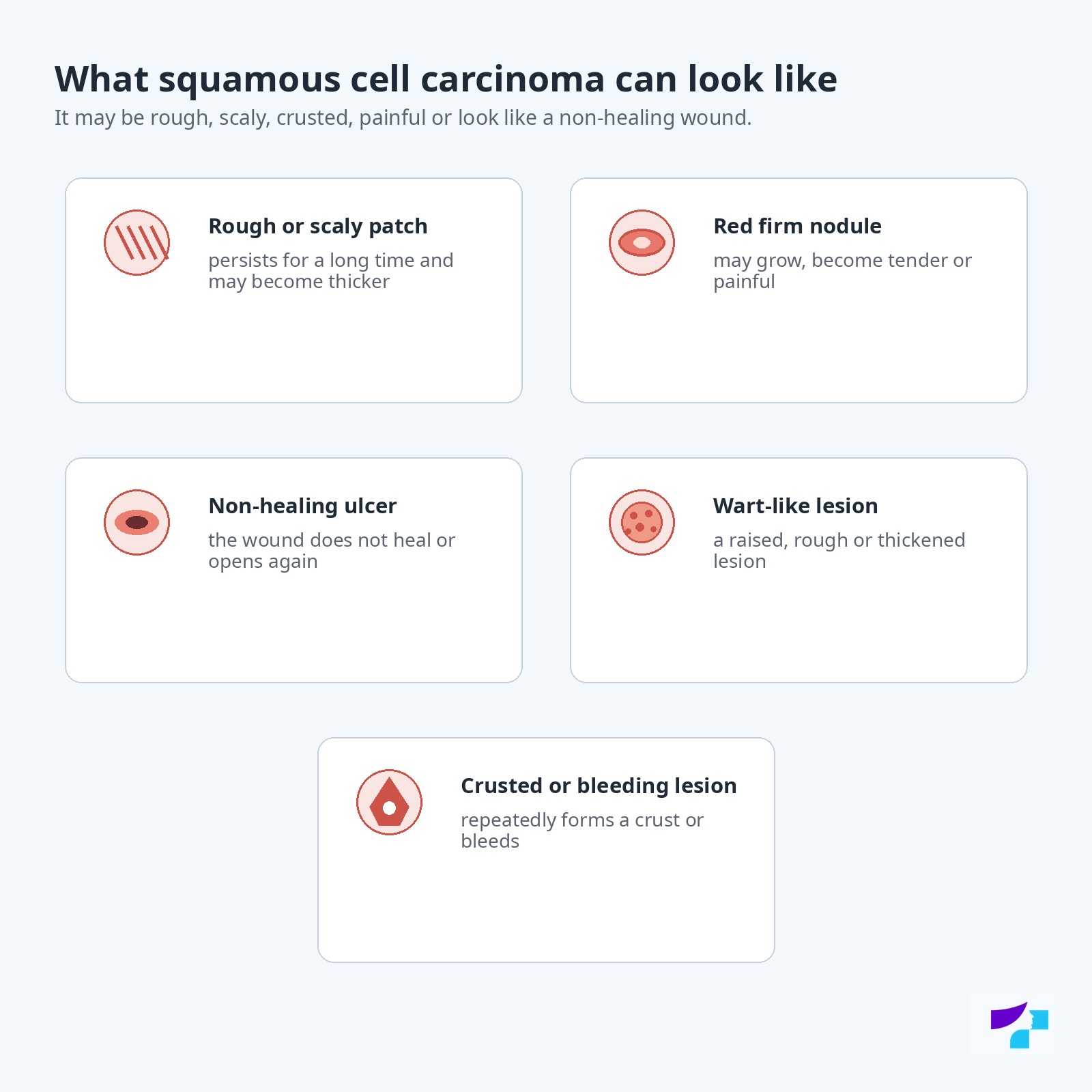
Typical appearances include:
- a rough or scaly patch;
- a red, firm nodule;
- a non-healing ulcer or wound;
- a wart-like growth;
- a thickened area of skin with a crust;
- a bleeding or painful skin lesion;
- a dry or rough patch on the lip;
- an ulcer or thickening in a scar or chronic wound.
At first, squamous cell carcinoma may look similar to actinic keratosis — a rough, sun-damaged patch. However, if the patch becomes thicker, painful, bleeds, ulcerates, oozes, grows quickly or looks like a nodule, it should be examined by a dermatologist.
A skin lesion is especially suspicious if it becomes thicker, more painful, starts oozing, ulcerating, bleeding or growing faster within a relatively short period of time. Squamous cell carcinoma does not always develop slowly and unnoticed — in some cases, a patient notices that a previously small rough patch or crust has quickly become a raised, tender or bleeding lesion.
Is squamous cell carcinoma always dark?
No. Squamous cell carcinoma is usually not dark like melanoma. It is more often pink, red, skin-coloured, yellowish and crusted, scaly or ulcerated. Dark colour is not the main sign. It is much more important that the lesion does not go away, grows, becomes thicker, bleeds, hurts, ulcerates or forms a crust.
Does squamous cell carcinoma hurt, itch or bleed?
Squamous cell carcinoma may not hurt, especially in the beginning. However, compared with basal cell carcinoma, it is more likely to become tender, painful, inflamed or bleeding. It may also itch, sting, ooze or cause discomfort.
Pain, bleeding, oozing or crusting are not the only signs, but they are important warning signals, especially if the lesion is located on the lip, ear, scalp, hand, finger, foot, scar or long-term sun-damaged skin.
Three to five main signs that may suggest squamous cell carcinoma
Squamous cell carcinoma is most often noticed through practical changes on the skin. The following signs are especially important.
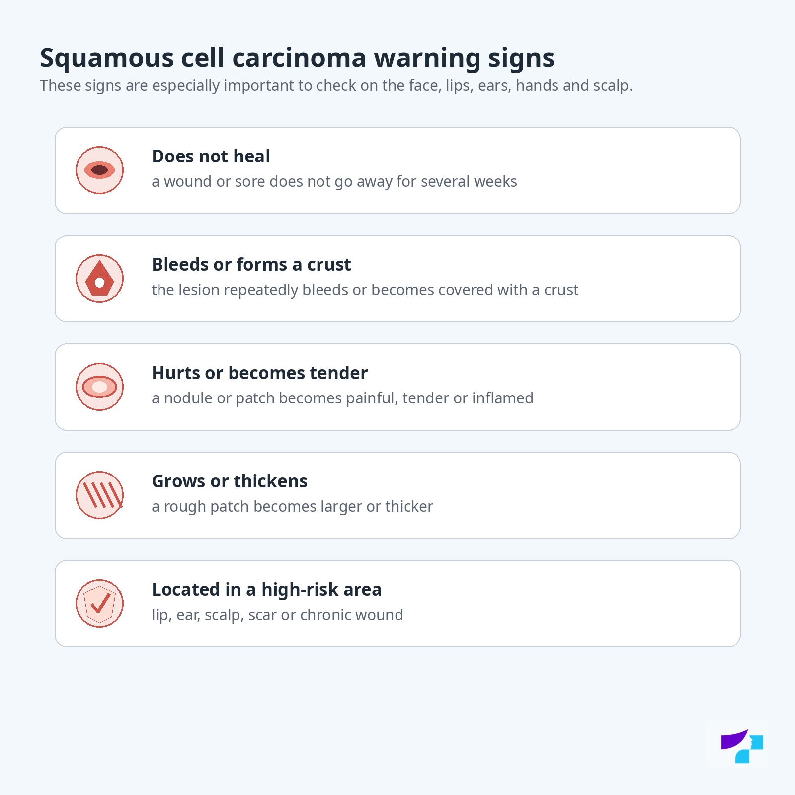
A rough or scaly patch that does not go away
If there is a rough, dry or scaly patch on sun-damaged skin that persists for a long time, becomes thicker or changes, it should be assessed by a dermatologist.
A non-healing ulcer or wound
A wound that does not heal for several weeks, opens again, bleeds, oozes or becomes covered with a crust may be a sign of squamous cell carcinoma.
A red, firm or painful nodule
Squamous cell carcinoma may look like a raised, firm, pink or red nodule that grows or becomes tender.
A wart-like lesion
Sometimes the tumour can resemble a wart — it may be raised, rough, thickened or have a crusted surface.
Bleeding, pain or crusting
If a lesion repeatedly bleeds, hurts, itches, becomes tender or forms a crust, it should not be observed at home for a long time.
Actinic keratosis, Bowen’s disease and squamous cell carcinoma
Squamous cell carcinoma is often linked to sun-damaged skin and precancerous changes. Two especially important terms are actinic keratosis and Bowen’s disease.
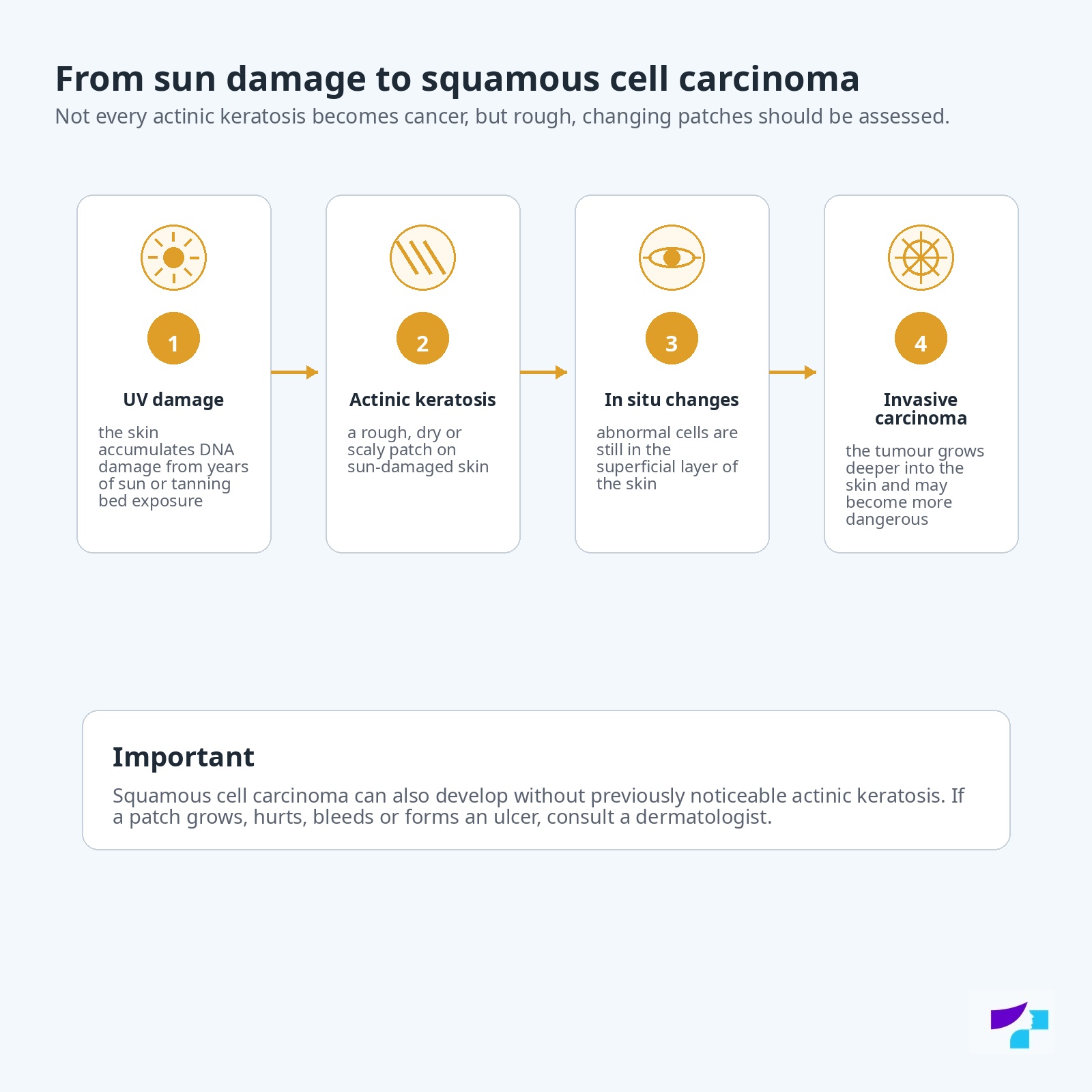
What is actinic keratosis?
Actinic keratosis is a sun-damaged skin change that usually looks like a rough, dry, scaly or sandpaper-like patch. It often appears on the face, ears, scalp, lips, backs of the hands, forearms and other sun-exposed areas.
Actinic keratosis itself is not yet an invasive skin cancer, but it is considered a precancerous condition. This means that some of these lesions can progress to squamous cell carcinoma, especially if they become thicker, painful, bleeding, oozing or ulcerated.
If there are many actinic keratoses on the skin, this may indicate broader sun damage in that area of skin. This situation is sometimes called field cancerisation — meaning that the risk is not only in one visible patch, but in the entire UV-damaged skin field. Therefore, a dermatologist may recommend treating not only a single lesion, but also a wider area of sun-damaged skin.
For a related topic, see the article Actinic keratosis.
Can actinic keratosis turn into squamous cell carcinoma?
Yes, actinic keratosis can turn into squamous cell carcinoma, but this does not happen with every lesion. What matters practically for the patient is not trying to predict the risk at home, but showing a dermatologist rough, changing, painful, bleeding, oozing or thickening patches.
Actinic keratoses on the lip, ears, scalp, backs of the hands and areas with extensive sun damage should be assessed especially carefully.
What is Bowen’s disease?
Bowen’s disease is squamous cell carcinoma in situ. This means that the malignant cells are located in the upper layer of the skin and have not yet grown deeper. It may look like a red, scaly, slowly growing patch that can resemble eczema, psoriasis or a fungal infection.
Bowen’s disease should be treated because some cases may become invasive. The treatment method is chosen by a doctor based on the location, size, number of lesions and the patient’s health status.
When should a rough patch be shown to a dermatologist?
A rough patch should be shown to a dermatologist if it:
- does not go away;
- becomes thicker;
- hurts or becomes tender;
- bleeds;
- oozes;
- forms a crust;
- ulcerates;
- enlarges quickly;
- is located on the lip, ear, scalp, backs of the hands, fingers, feet or in a scar;
- appears in a person with weakened immunity;
- is one of many sun-damaged patches.
Where does squamous cell carcinoma most often develop?
Squamous cell carcinoma most often develops in sun-damaged areas. Common locations include:
- the face;
- the lower lip;
- the ears;
- the nose;
- the scalp, especially in people with thinner hair or baldness;
- the neck;
- the backs of the hands;
- the forearms;
- the shoulders;
- the lower legs.
However, squamous cell carcinoma can also develop in areas that are not regularly exposed to the sun, especially where there is long-term inflammation, a chronic wound, a scar, a burn scar, an ulcer or risk related to immunosuppression.
Lesions on the lip, ear, scalp, fingers, toes, palms, soles, scars and chronic wounds should be assessed particularly carefully. In these locations, lesions may be noticed later or be associated with more complex treatment.
Can squamous cell carcinoma occur on the lip?
Yes. Squamous cell carcinoma can develop on the lip, especially the lower lip, which often receives sun exposure. Warning signs may include roughness, long-lasting dryness, a crack, ulcer, crust, bleeding, thickening or soreness on the lip.
The lip is an important location because squamous cell carcinoma in this area may be higher risk than many superficial skin lesions. Therefore, persistent roughness or an ulcer on the lip should not be treated only as “dry lips”.
Can squamous cell carcinoma occur on the ear or scalp?
Yes. The ear and scalp are important locations. On the ear, squamous cell carcinoma may look like a painful, crusted, bleeding or rough lesion. On the scalp, the lesion can be harder to notice, especially if a person does not check this area themselves.
Lesions in these areas should not be ignored for a long time, because diagnosis may be delayed and treatment may become more complicated.
Can squamous cell carcinoma occur on the fingers, feet or palms?
Yes. Squamous cell carcinoma can also develop on the fingers, toes, palms and soles. In these areas it may be mistaken for a wart, injury, ingrown nail, chronic fissure or non-healing wound.
If there is a lesion on a finger, foot or palm that grows, hurts, bleeds, thickens, does not heal or repeatedly forms a crust, it should not be treated for a long time as a wart or injury without a doctor’s assessment.
Can squamous cell carcinoma develop in a scar or chronic wound?
Yes. Squamous cell carcinoma can develop in chronic wounds, ulcers, burn scars or areas of long-term inflammation. These tumours are sometimes described as carcinomas developing on the background of chronic damage. They may be higher risk than some sun-damage-related tumours.
If a new thickening, ulceration, bleeding, pain, unpleasant smell, oozing or growing lesion appears in the area of a scar, ulcer or chronic wound, a medical examination is needed.
Can squamous cell carcinoma occur in the mouth or genital area?
Yes. Squamous cell carcinoma can also develop on mucous membranes, for example in the mouth or genital area. These cases may differ from classic sun-induced cutaneous squamous cell carcinoma in terms of risk factors, diagnosis and treatment. In certain locations, HPV infection, smoking, chronic irritation or other factors may be important.
This article mainly describes cutaneous squamous cell carcinoma. If the lesion is located in the mouth, throat, genital area or other mucous membranes, the diagnostic and treatment pathway may differ, and the involvement of an appropriate specialist is needed.
In the mouth, warning signs may include a non-healing ulcer, a white or red patch, thickening, pain or bleeding. In the genital area, suspicious findings include non-healing, ulcerated, thickened or bleeding lesions. In such cases, the appropriate specialist should be consulted — a dermatologist, venereologist, gynaecologist, urologist, dentist or oncologist, depending on the location.
Why does squamous cell carcinoma develop?
Squamous cell carcinoma develops when DNA damage accumulates in skin cells and the cells begin to divide uncontrollably. The main cause of this damage is ultraviolet radiation from the sun and tanning beds. UV radiation damages the genetic material of cells, and over time these changes can lead to precancerous changes and tumour development.
Squamous cell carcinoma is not an infection and is not contagious. It does not pass from one person to another through touch, shared towels, swimming pools or everyday contact.
In certain locations or specific situations, the risk of squamous cell carcinoma can also be increased by chronic inflammation, scars, long-standing wounds, weakened immunity, certain chemicals, ionising radiation, HPV infection and smoking.
Development mechanism of squamous cell carcinoma
In the case of squamous cell carcinoma, it is more accurate to talk about a development mechanism rather than a mechanism of infection, because it is not a contagious infectious disease.
In simple terms, development can be described as follows:
- UV radiation or other damaging factors affect the DNA of skin cells;
- the body repairs some of the damage, but some remains;
- precancerous changes, such as actinic keratosis, may appear in the skin;
- cells may become increasingly atypical and lose normal growth control;
- squamous cell carcinoma in situ or invasive squamous cell carcinoma develops;
- an invasive tumour can grow deeper into the skin and, in certain cases, spread to lymph nodes.
This process is not the same in every patient. In some people, lesions remain as rough patches for years, while in others changes may progress faster, especially in the setting of weakened immunity or a high-risk tumour.
Timeline of squamous cell carcinoma development
The development of squamous cell carcinoma can vary, but it can be explained to patients in several stages.
UV damage and skin ageing
Over the years, the sun or tanning beds damage skin cells. The skin may become rougher, more blotchy, thinner, with pigment changes and small blood vessels.
Actinic keratosis
Rough, dry, scaly patches appear on sun-damaged skin. These may be precancerous lesions. Not every such patch becomes cancer, but they should be assessed.
Squamous cell carcinoma in situ
Abnormal cells are located in the upper layer of the skin. This condition may be called Bowen’s disease. It is not yet a tumour that has grown deeply, but it should be treated.
Invasive squamous cell carcinoma
The tumour grows deeper into the skin. It may become painful, thickened, ulcerated, oozing, bleeding and enlarged.
Risk of spreading
In high-risk cases, the tumour can spread to lymph nodes or other parts of the body. Therefore, timely diagnosis and treatment are essential.
Who has a higher risk of developing squamous cell carcinoma?
Squamous cell carcinoma can develop in anyone, but the risk is higher in certain groups.
The risk is increased in people who have:
- fair skin, blond or red hair, light-coloured eyes;
- skin that burns easily and tans poorly;
- long-term or repeated sun exposure;
- tanning bed use;
- many actinic keratoses;
- a previous skin cancer;
- weakened immunity;
- organ transplantation and immunosuppressive therapy;
- chronic wounds, ulcers or scars;
- previous radiotherapy in a certain skin area;
- long-term contact with certain chemicals;
- smoking, especially in the context of lip and oral cavity risk;
- certain HPV infections, especially in mucosal and genital areas;
- older age, because skin damage accumulates over a lifetime.
Fair skin increases the risk because it contains less melanin — the pigment that partially protects against UV radiation. However, squamous cell carcinoma can also occur in people with darker skin, especially in scars, chronic wounds or areas that are not regularly checked.
Is the risk higher after organ transplantation?
Yes. In people after organ transplantation and in people receiving long-term immunosuppressive therapy, the risk of squamous cell carcinoma is significantly higher. The immune system helps the body control atypical cells and viral infections. When immune function is suppressed, the risk and aggressiveness of skin cancers can increase.
For these patients, regular dermatologist check-ups, sun protection and rapid response to new or changing skin lesions are especially important.
Can squamous cell carcinoma be hereditary?
Most squamous cell carcinomas are not directly hereditary. However, a person can inherit skin type, sensitivity to the sun or rare genetic diseases that increase skin cancer risk. Family history may be important if skin cancer occurs frequently in the family or at a young age.
Does the risk of another skin cancer increase after one squamous cell carcinoma?
Yes. If a person has already had squamous cell carcinoma, they have a higher risk of developing another squamous cell carcinoma, basal cell carcinoma or another skin cancer in the future. This is related to shared risk factors — accumulated UV damage, skin type, immunity and individual predisposition.
Therefore, regular skin follow-up is important after treatment.
When should you see a dermatologist?
You should see a dermatologist if there is a lesion on the skin or mucous membrane that:
- does not go away;
- grows or thickens;
- is rough or scaly;
- bleeds;
- oozes;
- hurts or becomes tender;
- repeatedly forms a crust;
- looks like a non-healing ulcer;
- looks like a wart but grows or bleeds;
- is located on the lip, ear, face, scalp, hand, finger, foot, scar or chronic wound;
- appears in a person with weakened immunity;
- appears in a place where there has already been skin cancer.
You should not wait until the lesion becomes very painful. Squamous cell carcinoma can be treatable when detected early, but delayed diagnosis increases treatment complexity.
How is squamous cell carcinoma diagnosed?
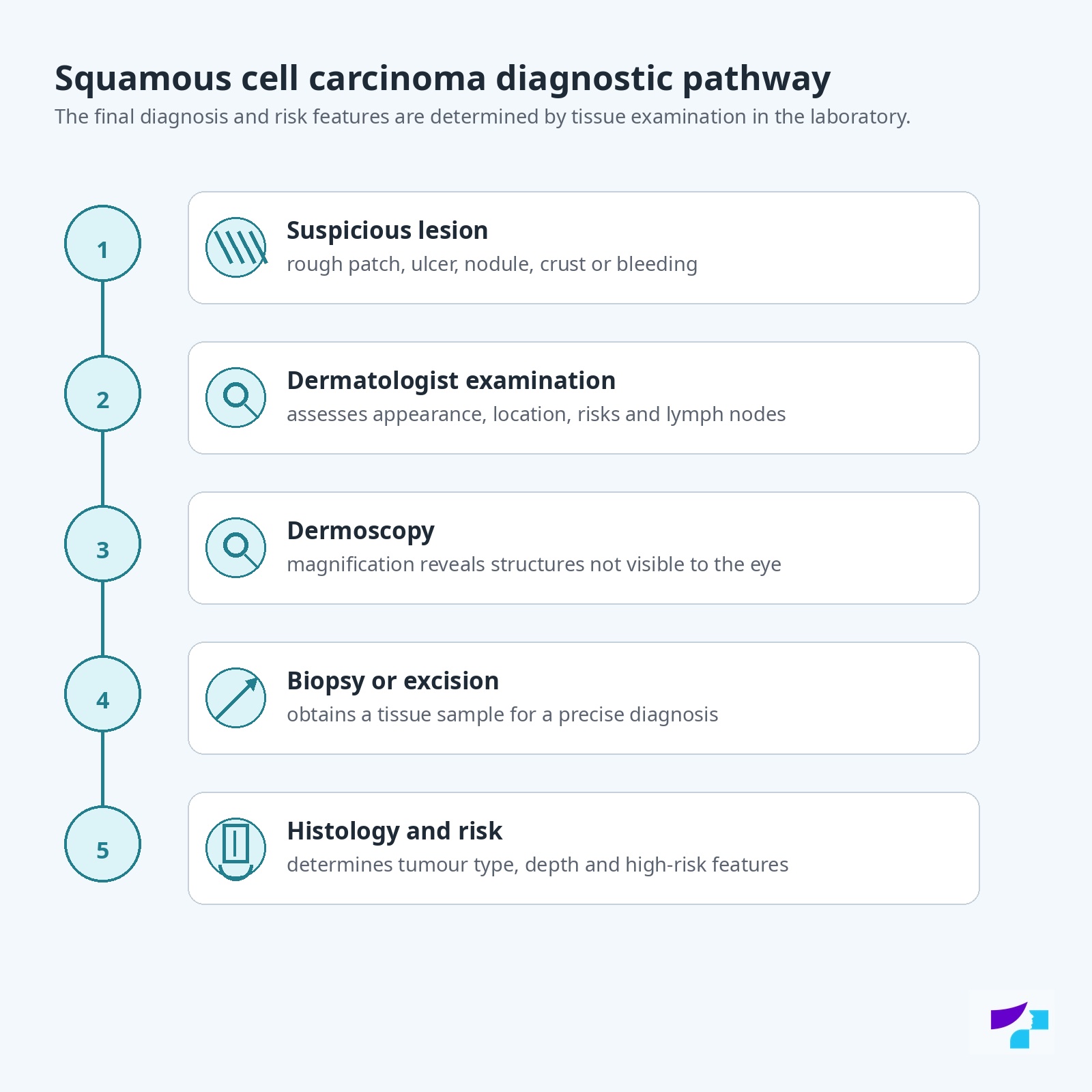
Diagnosis begins with a dermatologist’s examination. The doctor assesses the lesion’s appearance, size, location, surface, crusting, ulceration, bleeding, oozing, pain, growth rate and risk factors. If the lesion is on the lip, ear, scalp, finger, foot, in a scar, chronic wound or the patient has weakened immunity, the doctor assesses high-risk features especially carefully.
Dermoscopy is often performed. Dermoscopy is examination of a skin lesion with an optical device called a dermatoscope. It allows structures that are not visible to the naked eye to be seen under magnification. Dermoscopy helps distinguish squamous cell carcinoma from actinic keratosis, basal cell carcinoma, a wart, inflammation or other skin lesions, but histology is often needed for the final diagnosis.
For related topics, see the articles Dermoscopy and Skin lesion examination.
When is a biopsy needed?
A biopsy means taking a tissue sample for microscopic examination. It is needed if the lesion is suspicious for squamous cell carcinoma or if the diagnosis cannot be safely established by examination and dermoscopy alone.
A biopsy helps determine:
- whether the lesion is squamous cell carcinoma;
- whether it is in situ or invasive;
- how deeply the tumour grows;
- whether high-risk histological features are present;
- which treatment method should be chosen.
In some cases, the doctor may recommend removing the entire lesion at once and sending it for histological examination.
What is histological examination?
Histological examination is the microscopic assessment of tissue. It is important because squamous cell carcinoma can resemble actinic keratosis, basal cell carcinoma, a wart, a chronic wound, inflammation or another skin lesion.
Histology helps determine the tumour type, depth, differentiation, invasion, margins and other features that influence the treatment plan and the risk of recurrence or metastasis.
When should lymph nodes be examined?
Lymph nodes should be assessed if squamous cell carcinoma is high-risk or if there are clinical signs that may suggest spread. The doctor may examine the nearest lymph nodes by palpation, but if needed, ultrasound, imaging or other investigations may be recommended.
Lymph node assessment may be especially important if the tumour is large, deep, recurrent, located on the lip, ear, scalp, in a scar or chronic wound, if the patient has weakened immunity, or if high-risk features are found on histology.
Can squamous cell carcinoma be diagnosed from a photo alone?
No. A photograph may help understand whether a lesion looks suspicious, but it does not replace a doctor’s examination, dermoscopy and histological examination. Squamous cell carcinoma can look similar to actinic keratosis, a wart, basal cell carcinoma, eczema, an ulcer or inflammation.
Therefore, a suspicious lesion should not be assessed only by internet images or an artificial intelligence answer.
What can squamous cell carcinoma be confused with?
Squamous cell carcinoma can be difficult to recognise, especially in the beginning. It can be confused with:
- actinic keratosis;
- Bowen’s disease;
- basal cell carcinoma;
- a wart;
- seborrhoeic keratosis;
- eczema;
- psoriasis;
- a fungal skin infection;
- a chronic wound;
- an inflamed spot;
- a scar;
- a trauma-related crust;
- melanoma, especially in atypical or inflamed lesions.
If the lesion does not go away, grows, bleeds, oozes, hurts, thickens or forms an ulcer, a dermatologist’s examination is needed.
How is squamous cell carcinoma treated?
Treatment of squamous cell carcinoma depends on tumour size, depth, location, histological features, spread, recurrence risk and the patient’s general health. The aim of treatment is to completely remove or destroy the tumour, reduce the risk of recurrence, preserve function and achieve the best possible aesthetic result.
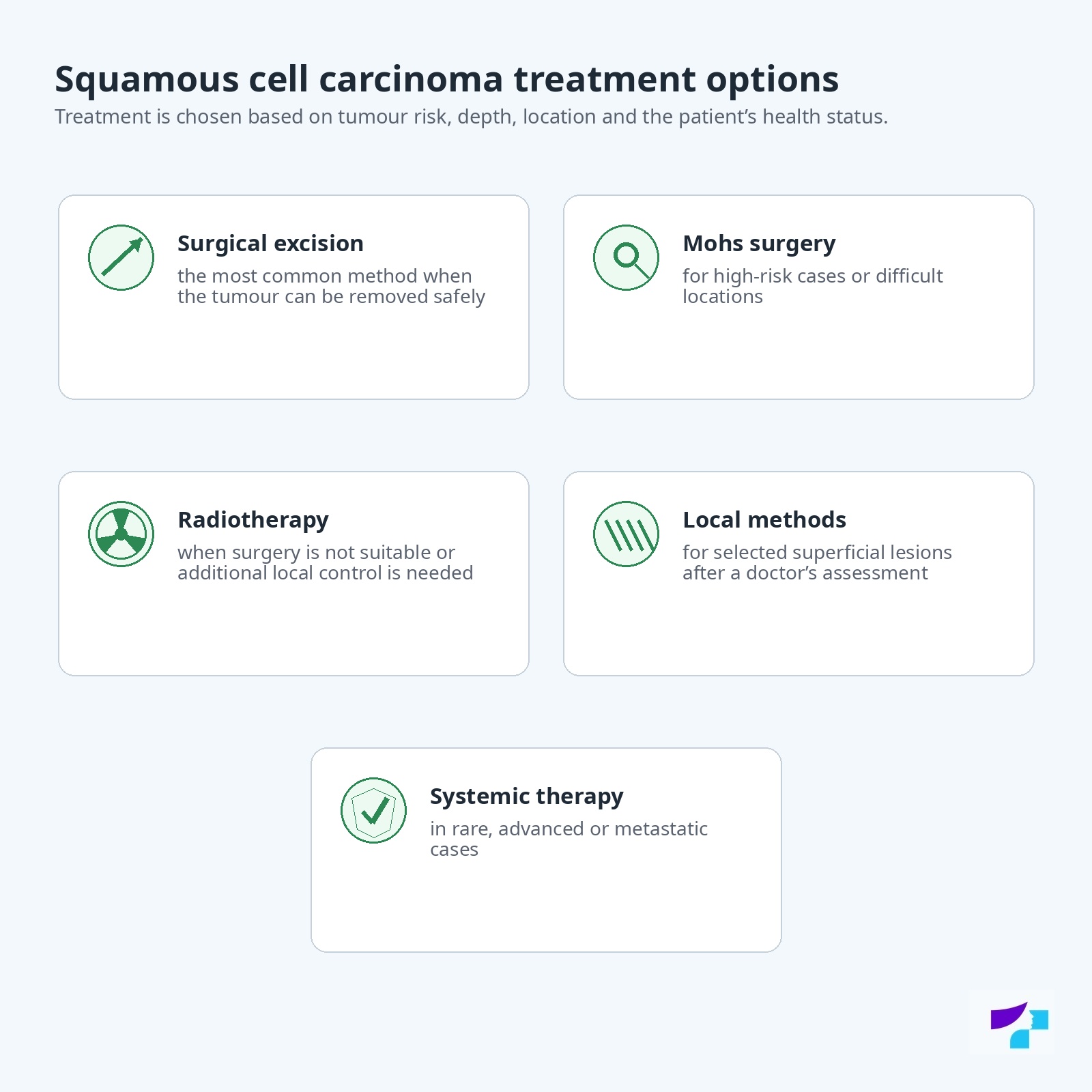
Main treatment options include:
- surgical excision;
- Mohs micrographic surgery or other micrographically controlled surgery in selected cases;
- radiotherapy, if surgery is not suitable or additional local control is needed;
- curettage with electrocautery in selected low-risk cases;
- cryotherapy or photodynamic therapy for certain superficial or precancerous changes;
- doctor-prescribed topical therapy for selected in situ lesions;
- systemic therapy in advanced, locally advanced or metastatic cases.
In practice, treatment can be divided into low-risk and high-risk situations. A small, superficial, clearly defined lesion in a low-risk location may require simpler treatment. A large, deep, painful, recurrent, fast-growing tumour, or a tumour on the lip, ear, scalp, in a scar, chronic wound or in an immunosuppressed patient, should be treated more carefully, often with lymph node assessment and involvement of a surgeon or oncologist.
This article does not list specific prescription medicines. Treatment may only be determined by a doctor after diagnosis, histology and individual risk assessment.
Can squamous cell carcinoma be cured?
Yes, squamous cell carcinoma that is diagnosed early and treated completely can often be cured. The prognosis is better if the tumour is small, superficial, has not spread and is treated in time.
However, delaying treatment can increase tumour depth, recurrence risk, the extent of scarring and the possibility of spread. Tumours on the lip, ear, scalp, fingers, feet, scars, chronic wounds and in people with weakened immunity should be treated especially carefully.
Is surgery always needed to treat squamous cell carcinoma?
Not always, but surgical excision is one of the main and most commonly used methods for treating invasive squamous cell carcinoma. Surgery allows the tumour to be removed and the tissue sent for histological examination to confirm the diagnosis and assess the margins.
For superficial or in situ lesions, in selected situations a doctor may choose other methods, such as topical therapy, photodynamic therapy or other procedures. However, in invasive, high-risk or deeper squamous cell carcinoma, treatment must be sufficiently radical to reduce the risk of recurrence and spread.
How is squamous cell carcinoma excised?
During surgical excision, the doctor removes the tumour together with a certain safety margin of healthy tissue. This safety margin is needed to reduce the risk that tumour cells remain in the skin. After excision, the tissue is sent for histological examination.
The extent of excision depends on the tumour size, depth, location and risk. In the face, lip, ear or scalp area, treatment planning may be more complex because both safe tumour removal and function and cosmetic outcome must be considered.
When is Mohs surgery needed?
Mohs micrographic surgery is a method in which the tumour is removed step by step, with the tissue margins checked under a microscope during treatment. This allows precise control of tumour removal and preserves more healthy skin.
Mohs surgery may be especially important for:
- tumours on the face, lip, ear, eyelid or nose;
- recurrent squamous cell carcinomas;
- tumours with unclear borders;
- high-risk tumours;
- locations where preserving as much healthy tissue as possible is important.
The availability and suitability of this method are assessed by a doctor.
When is radiotherapy used?
Radiotherapy may be used when surgery is not suitable, when the tumour is in a complex location, when the patient’s health does not allow surgery, or when additional local control is needed after treatment. Radiotherapy may also be used in selected cases when the tumour has spread or there is a high risk of recurrence.
The decision about radiotherapy is made by a specialist team after assessing tumour and patient factors.
Can squamous cell carcinoma be treated with cream?
Some superficial lesions or squamous cell carcinoma in situ can, in certain situations, be treated with doctor-prescribed topical prescription therapy. However, invasive squamous cell carcinoma usually cannot be treated safely with cream alone.
Self-treatment with over-the-counter products, acids, folk remedies or products recommended online is not safe and can delay diagnosis.
Can squamous cell carcinoma be treated with laser?
Laser is not a universal or standard method for treating invasive squamous cell carcinoma. A suspicious lesion must not simply be “removed with laser” without a diagnosis and treatment plan, because tissue histology may be needed for diagnosis, risk assessment and treatment planning.
Can squamous cell carcinoma be treated with cryotherapy or photodynamic therapy?
Cryotherapy and photodynamic therapy may be suitable for selected superficial lesions or precancerous changes, such as actinic keratosis or some in situ lesions. They are not suitable for all squamous cell carcinomas, especially if the tumour is invasive, deep, painful, high-risk or located in an important area.
The method is chosen by a doctor after diagnosis and risk assessment.
When is systemic therapy needed?
Systemic therapy means treatment that acts on the whole body. It may be needed in rare cases when squamous cell carcinoma is locally very advanced, cannot be treated completely with local methods, or has spread to lymph nodes or other organs.
In such cases, an oncologist is involved in treatment. The specific type of therapy is chosen according to tumour spread, histology, the patient’s health status and other factors.
When is an oncologist involved in treatment?
An oncologist is involved if squamous cell carcinoma is high-risk, advanced, recurrent, affects lymph nodes, cannot be completely removed with simple local treatment, or if radiotherapy or systemic therapy is needed.
Sometimes the treatment plan is developed by a multidisciplinary team — a dermatologist, surgeon, oncologist, radiation therapist, pathologist and other specialists.
Does a scar remain after squamous cell carcinoma removal?
After surgical excision, a scar usually remains. The appearance of the scar depends on the tumour size, location, extent of excision, individual wound healing and post-operative care.
In the face, lip, ear or scalp area, the doctor must plan not only safe tumour removal, but also preservation of function and aesthetic outcome. However, the main goal is always complete and safe tumour treatment.
Can squamous cell carcinoma be treated at home?
No. Squamous cell carcinoma must not be treated at home. Self-treatment can delay diagnosis, cause infection, scarring, bleeding or allow the tumour to continue growing.
A suspicious lesion should not be burned, cut, scratched, “dried out”, treated with acids or managed according to internet advice. If the lesion is non-healing, rough, bleeding, painful or growing, a dermatologist should be consulted.
What should not be done if a lesion could be squamous cell carcinoma?
A suspicious skin lesion should not be:
- burned with acids or folk remedies;
- cut or traumatised at home;
- treated for a long time as eczema if there is no improvement;
- removed with laser without a diagnosis;
- scratched off or “dried out”;
- assessed only by internet images;
- left untreated if it is located on the lip, ear, scalp, finger, foot, scar or chronic wound;
- ignored if the patient has weakened immunity.
A suspicious lesion should first be assessed by a doctor. If needed, dermoscopy, biopsy or excision with histological examination should be performed.
Is squamous cell carcinoma dangerous?
Yes, squamous cell carcinoma can be dangerous. Many cases are treatable if detected early, but squamous cell carcinoma has a higher risk of spreading than basal cell carcinoma. It can grow deeper into tissues, damage surrounding structures, recur after treatment and, in certain cases, spread to lymph nodes or other parts of the body.
Can squamous cell carcinoma metastasise?
Yes, squamous cell carcinoma can metastasise, although most early treated cases do not spread. The risk of metastasis is higher if the tumour is large, deep, poorly differentiated, recurrent, located on the lip, ear, scalp or in a scar, or if the patient has weakened immunity.
Most often, squamous cell carcinoma may first spread to the nearest lymph nodes.
What happens if squamous cell carcinoma is not treated?
Untreated squamous cell carcinoma can continue to grow, become deeper, more painful, bleed, ooze, ulcerate and damage surrounding tissues. It can lead to a larger scar and more complex treatment. In high-risk cases, it can spread to lymph nodes or elsewhere in the body.
Therefore, squamous cell carcinoma should not be postponed simply because the lesion seems “small” or is not very painful at first.
Can squamous cell carcinoma damage the lip, ear or nose?
Yes. If the tumour is located on the lip, ear, nose or other anatomically sensitive areas, it can grow deeper and damage tissues that have important functional and aesthetic significance. Treatment in these locations must be planned especially carefully.
Can squamous cell carcinoma return after excision?
Yes, squamous cell carcinoma can return, or recur. The risk of recurrence depends on the tumour’s risk features, location, histology, treatment method and whether the tumour was removed completely.
After treatment, regular follow-up is important because a person may develop not only recurrence, but also a new squamous cell carcinoma, basal cell carcinoma or another skin cancer.
Which squamous cell carcinoma cases are high-risk?
High-risk features may include:
- large tumour size;
- deep growth into tissues;
- rapid growth;
- recurrence after previous treatment;
- poor differentiation on histology;
- perineural invasion, when the tumour grows around nerves;
- location on the lip, ear, scalp, eyelid, genital area, fingers, feet, in a scar or chronic wound;
- weakened immunity;
- a tumour after organ transplantation;
- lymph node involvement.
In high-risk cases, treatment and follow-up are usually more intensive.
What is the prognosis of squamous cell carcinoma?
The prognosis is usually good when squamous cell carcinoma is detected early and treated completely. The prognosis becomes more serious if the tumour is large, deep, recurrent, poorly differentiated, located in a high-risk area or has spread to lymph nodes.
The prognosis is influenced by:
- tumour size;
- depth;
- histological type;
- location;
- immune status;
- recurrence;
- lymph node involvement;
- completeness of treatment;
- ongoing follow-up.
How can the risk of squamous cell carcinoma be reduced?
The risk of squamous cell carcinoma cannot be reduced to zero, but it can be lowered by limiting UV radiation and treating precancerous changes in time.
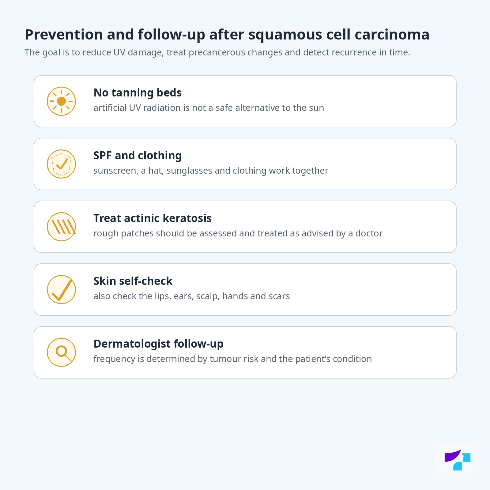
The most important prevention principles are:
- avoid sunburn;
- do not use tanning beds;
- seek shade during intense UV radiation;
- wear a hat, sunglasses and clothing that covers the skin;
- use a broad-spectrum sunscreen with appropriate SPF;
- check the skin regularly;
- treat actinic keratoses according to a doctor’s instructions;
- do not ignore non-healing wounds, scar changes or ulcers;
- monitor the skin especially carefully in people with weakened immunity;
- after skin cancer treatment, follow the dermatologist’s follow-up plan.
Does SPF help reduce the risk of squamous cell carcinoma?
When used correctly, SPF helps reduce skin damage caused by UV radiation. However, SPF is not the only protective measure and is not permission to stay in the sun longer. Combined protection works best: shade, clothing, a hat, sunglasses, sunscreen and avoiding tanning beds.
Should actinic keratosis be treated to reduce risk?
Yes, actinic keratosis should be assessed and treated according to a doctor’s instructions, especially if there are many lesions, they become thicker, hurt, bleed or are located in high-risk areas. Treating actinic keratosis can help reduce the risk of squamous cell carcinoma developing in that specific skin field.
If there are many actinic keratoses, the goal of treatment may be not only to remove a single patch, but also to control the entire sun-damaged skin field. This helps reduce the risk of new precancerous and cancerous lesions.
Can you sunbathe after a squamous cell carcinoma diagnosis?
Intentional sunbathing is not recommended. After a squamous cell carcinoma diagnosis, the skin should be protected especially carefully, because UV damage is one of the main risk factors and the person may have an increased risk of new skin cancers.
This does not mean that you cannot go outside, but sunburn, tanning beds and prolonged intense sun exposure should be avoided.
How often should the skin be checked after squamous cell carcinoma?
The frequency of follow-up is determined by the doctor. It depends on tumour risk, location, treatment method, histology, immune status and whether the patient has had other skin cancers.
High-risk patients usually need more frequent follow-up. It is important to check the skin regularly and contact a doctor promptly if a new, changing, bleeding, painful or non-healing lesion appears.
How should the skin be checked at home after treatment?
During skin self-checks, examine:
- the face, ears, lips and neck;
- the scalp;
- the backs of the hands and forearms;
- the shoulders, chest and back;
- the fingers, toes, feet and nails;
- scars and chronic wounds;
- the previously treated area;
- any areas where new roughness, an ulcer, crust, oozing or nodule appears.
For checking the back and scalp, help from a close person or a mirror may be useful. If a person has extensive sun damage, actinic keratoses or previous skin cancer, self-checks do not replace dermatologist follow-up.
What is well established in medicine at present?
It is medically well established that ultraviolet radiation is an important risk factor for squamous cell carcinoma. The link between actinic keratosis and some squamous cell carcinomas is also well established, as is the role of weakened immunity in increasing tumour risk and aggressiveness.
Evidence-based diagnosis includes a doctor’s examination, dermoscopy and histological examination if the lesion is suspicious. In treatment, complete removal or destruction of the tumour with an appropriate method is essential, taking tumour risk into account.
What is still being discussed and developed in medicine?
Medicine continues to improve the identification of high-risk squamous cell carcinoma in order to better understand which patients need more intensive treatment and follow-up. Digital dermoscopy, skin lesion photography, artificial intelligence support tools and personalised risk assessment are developing.
For advanced or metastatic squamous cell carcinoma, systemic treatment approaches continue to develop, including immunotherapy-type treatment in certain situations. Such treatment is not needed for most early squamous cell carcinomas, but it may be important in complex cases where the tumour cannot be fully treated with local methods.
References and authoritative sources
- Centre for Disease Prevention and Control of Latvia (SPKC): Don’t burn — choose to live
- Centre for Disease Prevention and Control of Latvia (SPKC): safe sun exposure
- US National Cancer Institute (NCI): skin cancer treatment — patient version
- US National Cancer Institute (NCI): skin cancer treatment — health professional version
- National Health Service (NHS): non-melanoma skin cancer — overview, symptoms and treatment
- National Health Service (NHS): treatment for non-melanoma skin cancer
- American Academy of Dermatology Association (AAD): guidelines for the management of cutaneous squamous cell carcinoma
- DermNet: cutaneous squamous cell carcinoma
- DermNet: actinic keratosis
- DermNet: intraepidermal squamous cell carcinoma / Bowen’s disease
- Cancer Research UK: squamous cell skin cancer
- European Association of Dermato-Oncology (EADO): European guidelines for skin cancer and melanoma diagnosis and treatment
Disclaimer
The information in this article is intended for informational and educational purposes and does not replace medical consultation, diagnosis or treatment. Self-assessment of skin and mucosal lesions may be wrong because squamous cell carcinoma can resemble actinic keratosis, a wart, eczema, a chronic wound, basal cell carcinoma, melanoma or other skin changes. If you notice a new, growing, bleeding, painful, itchy, crusted, oozing, non-healing or otherwise suspicious skin or mucosal lesion, consult a dermatologist, dermato-oncologist, surgeon, oncologist or an appropriate specialist according to the location, such as a dentist, gynaecologist or urologist. Do not try to treat, burn, cut, scratch or remove suspicious lesions on your own. If the lesion grows rapidly, bleeds significantly, ulcerates, becomes very painful, signs of infection appear, lymph nodes enlarge or general symptoms occur, urgent medical assessment is needed.
