Seborrheic keratosis is one of the most common benign skin lesions in adults. It may look like a brown, grey, yellowish or black skin growth with a rough, waxy or wart-like surface. Patients often describe it as a lesion that looks as if it is “stuck on” the skin.
In most cases, seborrheic keratosis is not dangerous and does not turn into skin cancer. However, patients cannot always reliably distinguish seborrheic keratosis from melanoma, a pigmented mole, basal cell carcinoma, actinic keratosis or another skin tumour. Any skin lesion that grows rapidly, bleeds, hurts, ulcerates, itches persistently or looks unusual should be shown to a dermatologist.
This article explains what seborrheic keratosis is, what it looks like, why it appears, when observation is sufficient, when a skin lesion check is needed and what removal options are available.
In Brief: Key Facts About Seborrheic Keratosis
- Seborrheic keratosis is usually a benign skin lesion.
- It appears more often in adulthood and becomes more common with age.
- The lesion may be skin-coloured, yellowish, grey, brown or almost black.
- It often looks like a “stuck-on” plaque or wart-like growth.
- It is not the same as seborrheic dermatitis.
- It is not a viral wart and is not contagious.
- If the diagnosis is clear and the lesion does not cause problems, treatment is often not needed.
- It can be removed if it rubs, itches, bleeds, becomes inflamed or causes cosmetic concern.
- Suspicious or changing skin lesions should be assessed with dermoscopy.
- Cutting, burning, freezing or treating the lesion with acids at home is not safe.
- If the diagnosis is uncertain, accurate diagnosis is more important than cosmetic removal.
What Is Seborrheic Keratosis?
Seborrheic keratosis is a benign lesion of the upper layers of the skin. It is sometimes called a “senile wart”, seborrheic wart or basal cell papilloma. The term “senile wart” is understandable for patients because these lesions become more common with age, but medically it can be misleading: seborrheic keratosis is not a true viral wart and is not infectious.
The word “seborrheic” also does not mean that the lesion arises from sebaceous glands or that it is related to seborrheic dermatitis. Seborrheic dermatitis is an inflammatory skin condition with redness and scaling, while seborrheic keratosis is a benign skin growth.
Seborrheic keratosis is usually not a precancerous condition. However, it may be dark, irregular, inflamed or crusted, and in such cases it can resemble more serious skin lesions. This is why accurate assessment by a dermatologist is important, rather than self-diagnosis based on online images.
In simple terms: seborrheic keratosis itself is usually not dangerous, but “a brown skin lesion” is not a diagnosis. First it is important to know what the lesion is, and only then can it be safely decided whether to leave it alone, monitor it or remove it.
What Does Seborrheic Keratosis Look Like?
Seborrheic keratosis can look very different from person to person. In some people it is light brown and almost flat; in others it is dark, raised, rough or wart-like. A typical feature is the “stuck-on” appearance: the lesion seems to sit on top of the skin rather than grow deeply into it.
Seborrheic keratosis may be:
- round or oval;
- flat or raised;
- smooth, waxy, scaly or wart-like;
- a few millimetres or several centimetres in size;
- single or multiple;
- skin-coloured, yellowish, grey, light brown, dark brown or black.
The most common areas include the face, neck, chest, back, shoulders, scalp, the area under the breasts and skin folds. Seborrheic keratosis usually does not occur on the palms, soles or mucous membranes.
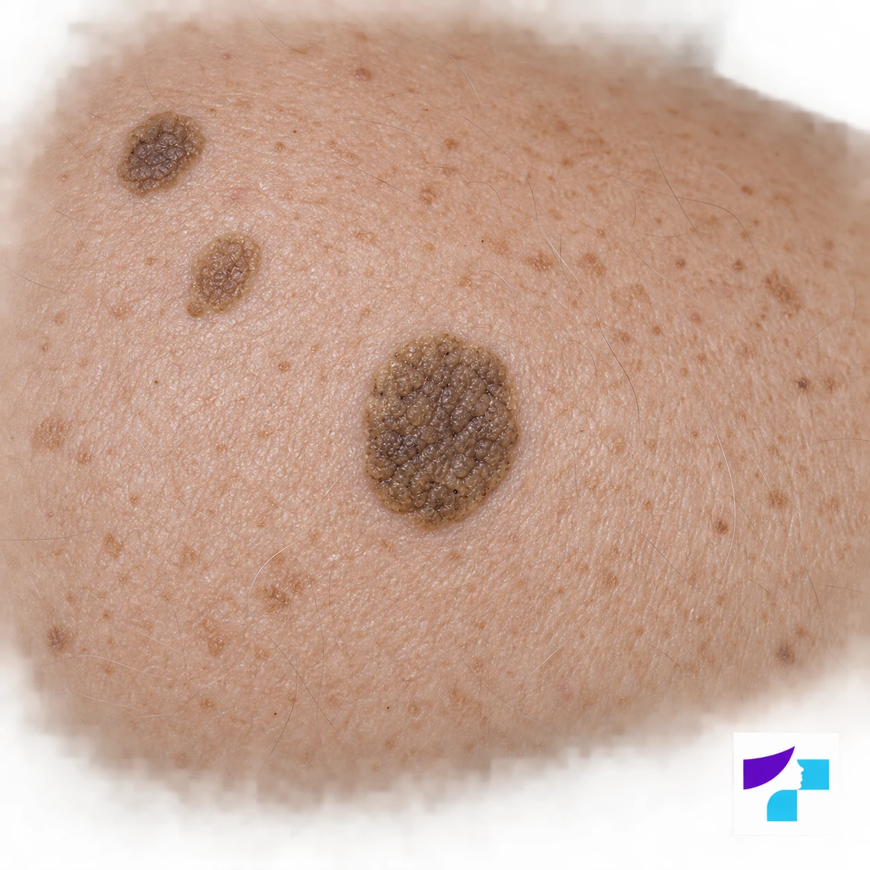
AI-generated illustration of the appearance of seborrheic keratosis: brown, raised lesions with a waxy, rough surface. This is not a photograph of a real patient; diagnosis requires assessment by a dermatologist.
| Feature | Typical for seborrheic keratosis | When to be cautious |
|---|---|---|
| Colour | Brown, grey, black, yellowish or skin-coloured | Very uneven pigmentation or sudden colour change |
| Surface | Waxy, rough, scaly or wart-like | Ulceration, bleeding or crusting without a clear reason |
| Shape | Round, oval and clearly demarcated | Asymmetry, blurred borders or rapid change in shape |
| Sensation | Usually painless; may itch or rub | Pain, persistent inflammation or rapid growth |
| Number | May be single or multiple | Sudden appearance of many lesions should be assessed by a doctor |
Why Does Seborrheic Keratosis Develop?
The exact cause of seborrheic keratosis is not fully known. It is clear that it becomes more common with age. Many people first notice lesions after the age of 40 or 50, although they may appear earlier.
Genetic predisposition may play a role: if several family members have many seborrheic keratoses, the likelihood of developing them may be higher. In some cases, sun exposure, skin ageing, individual skin characteristics and friction in skin folds or areas irritated by clothing may also contribute.
Seborrheic keratosis is not a sign of poor hygiene. It is not a fungal infection, not a viral wart and not contagious. It cannot be passed to other people and does not spread from one body area to another like an infection.
Is Seborrheic Keratosis Dangerous?
In most cases, seborrheic keratosis is not dangerous. It is benign and usually does not turn into cancer. The main risk is not the lesion itself, but the possibility of mistaking another, more serious skin lesion for seborrheic keratosis.
Melanoma, basal cell carcinoma, squamous cell carcinoma, actinic keratosis or an atypical mole may sometimes resemble seborrheic keratosis. Sometimes skin cancer may occur next to a benign lesion or look like an inflamed, bleeding or dark keratosis.
Therefore, if a lesion is new, grows quickly, changes colour, bleeds, ulcerates, hurts or looks different from other skin lesions, it should be shown to a dermatologist. The dermatologist may perform dermoscopy and, if uncertainty remains, recommend biopsy or removal with histological examination.
How to Distinguish Seborrheic Keratosis From Other Skin Lesions
Sometimes seborrheic keratosis is fairly easy to recognise, especially when it is typical: clearly demarcated, waxy, rough and “stuck on”. However, visual assessment alone is not always reliable because pigmented skin lesions can be misleading.
| Lesion | How it may look | Why it may be confused | What to do |
|---|---|---|---|
| Seborrheic keratosis | Stuck-on, waxy, brown, grey or black lesion | May be dark, rough and irregular | Dermatologist assessment, dermoscopy |
| Melanoma | Changing, pigmented, often asymmetric lesion | May resemble a dark keratosis | Prompt dermatologist assessment |
| Mole | Pigmented macule or raised lesion | May have a similar colour and size | Assess changes and perform dermoscopy |
| Actinic keratosis | Rough, scaly patch on sun-damaged skin | May be rough and crusted | Assess as a possible precancerous lesion |
| Viral wart | Rough, wart-like lesion | Surface may look similar | Medical assessment, especially in unusual locations |
| Basal cell carcinoma | Shiny, ulcerated, crusted or bleeding lesion | May resemble irritated keratosis | Dermatologist assessment and treatment plan |
At home, patients should pay particular attention to the ABCDE signs used when assessing pigmented lesions: asymmetry, border irregularity, multiple colours, increasing diameter and evolution or change over time. If any of these signs appear, a skin lesion check is needed.
It is especially important to distinguish seborrheic keratosis from actinic keratosis. Seborrheic keratosis is usually a benign, “stuck-on”, waxy or wart-like lesion, whereas actinic keratosis is a chronic change in sun-damaged skin that often feels like a rough, scaly or sandpaper-like patch and is considered a precancerous condition. A persistent rough patch on sun-exposed skin that thickens, hurts, bleeds or crusts should not be regarded simply as an “age wart”.
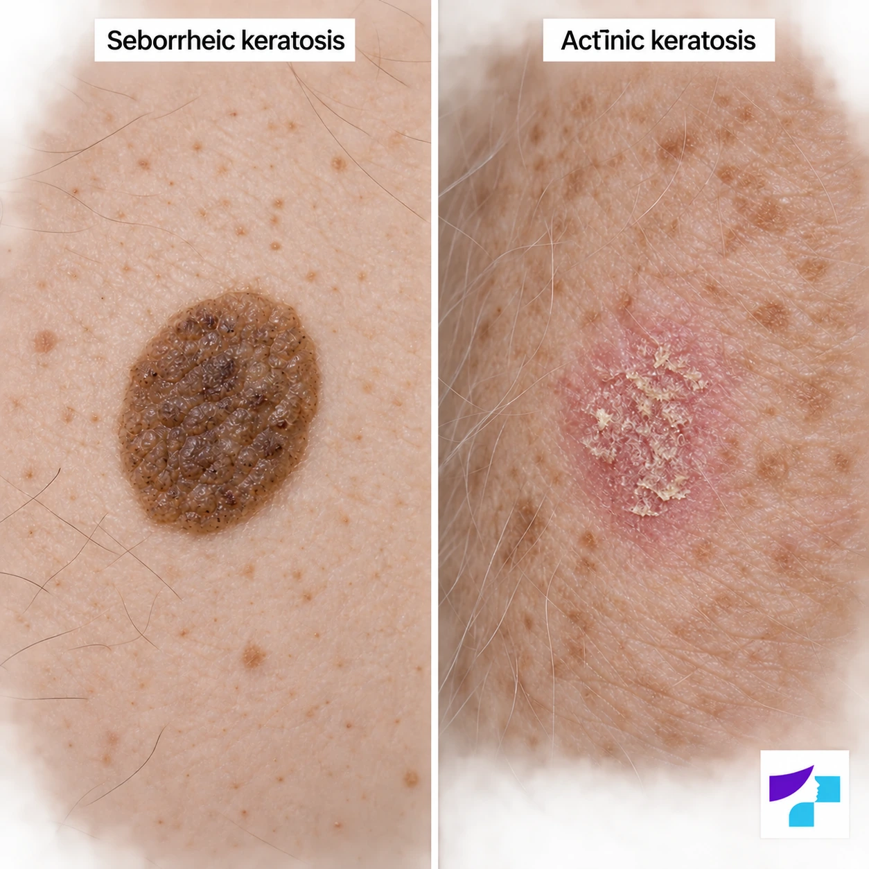
AI-generated illustrative comparison: a lesion resembling seborrheic keratosis on the left and a skin change resembling actinic keratosis on the right. Both conditions can vary in appearance; this illustration does not replace assessment by a dermatologist.
When Should You See a Dermatologist?
A dermatologist consultation is needed if:
- the lesion grows rapidly;
- its colour, shape or surface changes;
- it bleeds or ulcerates;
- pain appears;
- it persistently itches or becomes inflamed;
- the lesion is very dark or uneven;
- a new, unusual lesion has appeared;
- the lesion looks different from the others;
- the patient has previously had melanoma or another skin cancer;
- there is uncertainty whether it is really seborrheic keratosis.
It is better to check a skin lesion earlier rather than wait until symptoms become pronounced. This is especially important for patients with many pigmented lesions, fair skin, repeated sunburns or a family history of skin cancer.
How Is Seborrheic Keratosis Diagnosed?
Seborrheic keratosis is usually diagnosed by a dermatologist after examining the skin and assessing the appearance of the lesion. Dermoscopy is often used. It is a non-invasive method of examining skin lesions with a special optical device. A dermatoscope helps the doctor see structures that are not visible to the naked eye.
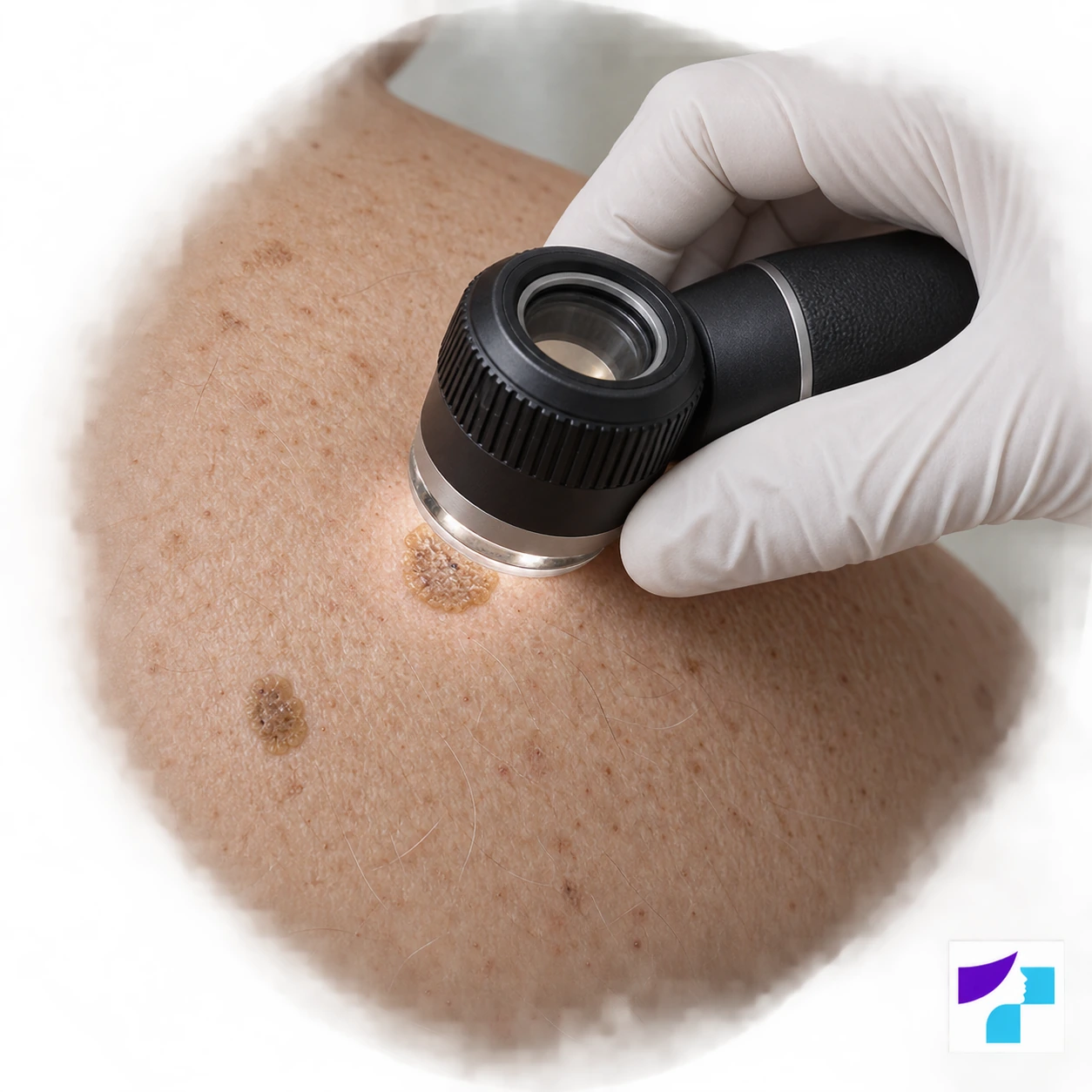
AI-generated illustration of a dermoscopy examination. A dermatoscope helps the dermatologist assess the structures of a skin lesion; this image does not document a real patient's examination.
During the consultation, the doctor may ask:
- when the lesion appeared;
- whether it has changed recently;
- whether there is itching, pain, bleeding or inflammation;
- whether it is traumatised by clothing, jewellery, shaving or combing;
- whether the patient has previously had skin tumours;
- whether there is a family history of melanoma or other skin cancers.
If the lesion looks atypical or the dermatologist is uncertain about the diagnosis, biopsy or complete removal with histological examination may be needed. Histology allows the tissue to be examined under a microscope and the diagnosis to be clarified.
Does Seborrheic Keratosis Need Treatment?
If the diagnosis is clear and the seborrheic keratosis does not cause problems, treatment is not mandatory. Many people live with these lesions for years, and medically they do not need to be removed simply because they are present.
Sometimes a drier or superficially scaly seborrheic keratosis may seem less rough or may partially flake off after bathing, sauna use or regular moisturising. This does not mean the lesion has been “cured”, and it does not replace dermatologist assessment if the lesion is dark, changing, bleeding or unclear.
Removal may be considered if the lesion:
- rubs against clothing or jewellery;
- is traumatised during shaving or combing;
- itches, bleeds or becomes inflamed;
- is in a visible area and causes cosmetic concern;
- creates diagnostic uncertainty;
- causes persistent anxiety for the patient.
Cosmetic removal is possible, but the diagnosis should be clear first. This is especially important for dark, pigmented or rapidly changing lesions.
Seborrheic Keratosis Removal Methods
The removal method should be chosen individually. It depends on the lesion’s size, thickness, colour, location, the patient’s skin type, the risk of scarring and whether histological examination is needed.
Cryotherapy With Liquid Nitrogen
Cryotherapy is a method in which the lesion is treated with liquid nitrogen. Cold damages the lesion cells; after the procedure redness, sometimes a blister or crust may form, and the keratosis gradually peels away.
This method may be suitable for thinner, clearly benign seborrheic keratoses. It is usually quick and does not require an incision, but it is not always the best choice for very thick, raised, dark or diagnostically unclear lesions. After cryotherapy, a lighter or darker patch may remain, especially in people with darker skin phototypes.
Curettage and Electrocoagulation
Curettage means gently removing the lesion with a special instrument, while electrocoagulation helps treat the tissue and stop minor bleeding. Local anaesthesia is usually used before the procedure.
This approach may be suitable for raised, rough and mechanically irritating lesions. After the procedure, a small wound forms and heals with a crust. Temporary redness, pigmentation change or a small scar may occur. If the lesion is suspicious, the doctor decides whether tissue should be sent for histology.
Laser Therapy
Seborrheic keratosis can be removed with a laser in selected cases, especially if the lesion is clearly benign and located in an aesthetically important area. A laser can precisely vaporise or remove a superficial lesion while reducing bleeding.
CO2 laser is one of the methods used in dermatology for removing clearly benign superficial skin lesions. Its advantage may be precision and a good cosmetic result, especially on the face or other visible areas. However, laser is not a universal method for all pigmented skin lesions.
If a lesion is dark, changing or diagnostically unclear, it should not simply be vaporised without assessment. In such cases, histological examination may be more important in order not to miss melanoma or another skin tumour.
Surgical or Shave Removal
Shave removal means that the lesion is shaved from the skin surface with a sharp instrument, usually under local anaesthesia. Surgical excision may be needed less often, for example if the lesion is atypical or a more complete tissue examination is required.
The advantage of these methods is the possibility to send tissue for histological examination. This is essential if there is even slight uncertainty about the diagnosis. In such cases, the cosmetic result still matters, but the first priority is to safely understand what the lesion is. Possible risks include bleeding, infection, scarring or pigmentation changes.
Topical Products and Home Methods
Seborrheic keratosis should not be cut, picked, burned, treated with acids or treated with unknown products at home. Home methods may cause burns, infection, scarring, pigmentation changes and prolonged inflammation. More importantly, self-removal may delay the diagnosis of skin cancer if the lesion is not actually seborrheic keratosis.
Over-the-counter products intended for warts or thickened skin are not a safe choice for pigmented lesions. The diagnosis should be known before any treatment is attempted.
Which Method Is Suitable in Which Situation?
| Situation | Possible approach | What can be achieved | Important caution |
|---|---|---|---|
| Small, clearly benign lesion | Cryotherapy, curettage, laser | Removal or reduction of the lesion | Possible pigmentation change |
| Lesion rubbing against clothing | Curettage, electrocoagulation, shave removal | Less irritation and discomfort | Wound care is important |
| Dark or atypical lesion | Dermoscopy, biopsy or histology | Accurate diagnosis | Assessment should not be delayed |
| Lesion on the face | Gentle method after assessment | Better cosmetic result | Scar and pigmentation risk should be considered |
| Multiple lesions | Individual plan | Removal of irritating or concerning lesions | Not all lesions need to be removed |
How Is the Most Suitable Removal Method Chosen?
Patients often think the main question is “laser or freezing”, but the dermatologist first assesses the diagnosis. If the lesion is a typical seborrheic keratosis, the method is chosen mainly according to cosmetic outcome, healing time, location and individual skin characteristics. If the lesion is atypical, safe diagnosis is the priority, not the fastest cosmetic removal.
The choice depends on several factors:
- whether the lesion is clinically and dermoscopically benign;
- whether histological examination is needed;
- whether the lesion is thin, thick, raised or flat;
- whether it is located on the face, scalp, back, chest or in a skin fold;
- how important the cosmetic result is to the patient;
- the risk of pigmentation changes and scarring;
- whether the patient can follow sun protection and aftercare instructions.
For example, cryotherapy may be a simple and quick method for thinner lesions, but it may not be the best option for very thick or very dark lesions. CO2 laser or another laser method may be suitable in aesthetically important areas if the diagnosis is clear. Shave removal or curettage is a practical option when the lesion is raised, rubs or tissue is needed for histology.
How to Care for the Skin After Removal
After the procedure, a small wound, redness or crust usually remains. Healing time depends on the method, lesion size and location, but often takes around one to two weeks. The doctor explains how to care for the skin in each specific case.
After seborrheic keratosis removal, it is usually important to:
- keep the area clean and dry as instructed by the doctor;
- not pick the crust;
- avoid aggressive cosmetic products on healing skin;
- avoid friction and trauma;
- use sun protection after healing, especially on the face and other exposed areas;
- temporarily avoid sauna, swimming pools, hot baths and intense heat if the doctor recommends it.
If increasing redness, pain, discharge, significant swelling or abnormal healing occurs, the doctor should be contacted.
Can Seborrheic Keratosis Be Prevented?
There is no reliable way to completely prevent seborrheic keratoses because age, genetic predisposition and individual skin characteristics play an important role. Sun protection is important for overall skin health and skin cancer prevention, but it does not guarantee that seborrheic keratoses will not appear.
In practice, it is advisable to examine the skin regularly, notice new or changing lesions and see a dermatologist in time. Regular skin checks are especially useful for people with many pigmented lesions, fair skin, repeated sunburns or a family history of skin cancer.
Many Seborrheic Keratoses Appeared Suddenly: What Does It Mean?
In many people, seborrheic keratoses gradually become more numerous with age, and this is usually a benign process. However, if many new lesions appear suddenly within a short time, especially together with itching, inflammation or general health symptoms, the situation should be assessed by a doctor.
A rare phenomenon has been described in medicine in which the sudden appearance of many seborrheic keratoses is associated with signs of internal disease. However, this association is not simple and is not always proven. The most important step for the patient is not to search for rare diagnoses independently, but to show the skin to a dermatologist so that the doctor can decide whether additional examination is needed.
What Happens During a Dermatologist Consultation?
During the consultation, the dermatologist examines the lesion and assesses whether it is consistent with seborrheic keratosis. Dermoscopy is often performed. If there are many lesions, the doctor may also assess other skin lesions to make sure there are no suspicious changes.
The consultation includes assessment of:
- the type and location of the lesion;
- whether it is benign or suspicious;
- whether observation, removal or histology is needed;
- which method is most suitable;
- the expected healing time;
- the risk of scarring or pigmentation change;
- how to care for the skin after the procedure.
This approach helps avoid a situation in which a lesion is removed cosmetically when it actually needed a more thorough diagnostic assessment.
How Much Does Seborrheic Keratosis Removal Cost?
The cost of removal is not the same for every case. It depends on the number, size and location of lesions, the chosen method, whether local anaesthesia is needed and whether the lesion must be sent for histological examination.
The price may also differ depending on whether the procedure is performed for medical reasons, for example because the lesion bleeds or is diagnostically unclear, or for cosmetic reasons. The exact cost can be determined during consultation or according to the clinic’s price list.
Can Seborrheic Keratosis Return After Removal?
If the seborrheic keratosis is completely removed, it usually does not return in the same place. However, people with a tendency to develop these lesions may develop new ones in other areas over time. This does not mean that the previous procedure was unsuccessful.
Some patients have many seborrheic keratoses that appear gradually over several years. In such cases, it is usually not necessary to remove all lesions. Priority is given to those that cause irritation, cosmetic concern or diagnostic uncertainty.
Risks and Side Effects
After seborrheic keratosis removal, temporary skin reactions may occur:
- redness;
- tenderness;
- crusting;
- mild swelling;
- bleeding;
- itching during healing.
Less commonly, infection, prolonged redness, a lighter or darker patch, a small scar or incomplete removal may occur. The risk of pigmentation changes is higher in darker skin phototypes and after more aggressive procedures.
A separate risk is diagnostic error if the lesion is removed or treated without adequate assessment. This is why it is important to make sure the lesion is truly benign before treatment.
What Should Not Be Done With Seborrheic Keratosis?
Seborrheic keratosis should not be:
- picked;
- scratched until bleeding;
- cut at home;
- burned;
- treated with acids;
- treated with unknown or aggressive products;
- frozen at home;
- diagnosed only from online images;
- removed cosmetically if it is suspicious;
- ignored if it changes.
If the lesion itches, bleeds or is repeatedly traumatised, it is safer to show it to a dermatologist and choose a controlled medical approach.
Common Myths About Seborrheic Keratosis
Myth: Seborrheic keratosis is always a wart.
In fact, it is not a viral wart and is not contagious.
Myth: Seborrheic keratosis is an infection.
It is not an infectious disease and cannot be passed from person to person.
Myth: It always needs to be removed.
If the diagnosis is clear and the lesion does not cause problems, treatment is often not needed.
Myth: If a lesion is brown, it must be a mole.
Brown skin lesions may be moles, seborrheic keratoses, lentigines or more serious skin tumours.
Myth: Melanoma always looks different from seborrheic keratosis.
Sometimes melanoma may resemble a benign lesion, which is why dermoscopy is important.
Myth: A skin lesion can be safely removed at home.
Self-removal may cause scarring, infection and delayed diagnosis.
Myth: After removal, lesions will never appear again.
The removed lesion usually does not return, but new seborrheic keratoses may appear elsewhere.
Myth: Laser is suitable for all pigmented lesions without examination.
A diagnosis is needed before laser treatment. Suspicious pigmented lesions should be assessed and sometimes sent for histology.
Summary
Seborrheic keratosis is a common and usually benign skin lesion that most often appears in adulthood. It may be light, dark, flat, raised, smooth or rough, which is why patients cannot always reliably distinguish it from other skin lesions.
If the lesion is stable, does not cause problems and the dermatologist confirms that it is benign, treatment is often not needed. If seborrheic keratosis rubs, itches, bleeds, is located in a cosmetically important area or creates diagnostic uncertainty, it can be removed using the most suitable method. The safest step is dermatologist consultation and dermoscopy, especially for dark or changing lesions.
References and Authoritative Sources
- American Academy of Dermatology: overview of seborrheic keratoses, symptoms and diagnosis
- American Academy of Dermatology: treatment and removal options for seborrheic keratoses
- DermNet: specialist dermatology information on seborrhoeic keratosis
- MedlinePlus Medical Encyclopedia: patient information on seborrheic keratosis
- British Association of Dermatologists: patient information on seborrhoeic keratosis
- Mayo Clinic: symptoms and causes of seborrheic keratosis
- Cleveland Clinic: causes, risks and treatment of seborrheic keratosis
