An epidermal or epidermoid cyst is a common, usually benign skin lesion. It is a closed capsule in or beneath the skin that is most often filled with keratin, a protein that forms skin, hair and nails. In everyday language, this lesion is often called a “sebaceous cyst”, “fatty cyst” or “atheroma”, but these terms are not always medically accurate because an epidermal cyst is generally not filled with fat.
An epidermal cyst often appears as a round, slowly growing lump beneath the skin. It may be painless, but can become red, warm, swollen and painful if it becomes inflamed, ruptures or is infected. A cyst should not be squeezed, punctured or emptied at home because this may worsen inflammation, promote infection and increase the risk of scarring, and it does not remove the cyst capsule.
What is an epidermal or epidermoid cyst?
The terms epidermal cyst and epidermoid cyst generally mean the same thing. Other terms used in medical literature include epidermal inclusion cyst, epidermoid cyst, infundibular cyst and keratin cyst.
It is a skin cyst that develops from cells of the epidermis, the outermost layer of the skin, or the upper part of a hair follicle. These cells continue to produce keratin, but instead of being shed from the skin surface, it accumulates inside a capsule. This creates a slowly growing nodule or lump beneath the skin.
An epidermal cyst is only one type of skin and subcutaneous cyst.
Epidermal cyst, epidermoid cyst, atheroma and sebaceous cyst: why are the terms confused?
People often use the terms “atheroma”, “sebaceous cyst” or “fatty cyst” to describe a lump beneath the skin containing whitish or yellowish material. An epidermal cyst, however, is generally filled with keratin and skin-cell debris rather than sebum from a sebaceous gland.
“Epidermal cyst” or “epidermoid cyst” is therefore more accurate. The word “atheroma” may be used informally as a broad description, but the diagnosis is made by a doctor after examination and, when needed, imaging or histological assessment.
| Term | What does it often mean in everyday use? | What is medically important? |
|---|---|---|
| Epidermal cyst | A skin cyst with a capsule | An accurate term for a common type of keratin-filled cyst |
| Epidermoid cyst | The same as an epidermal cyst | An alternative term |
| Atheroma | A lump beneath the skin or “fatty cyst” | A commonly used but not always accurate informal term |
| Sebaceous cyst | A “cyst filled with fat” | An epidermal cyst is generally not filled with fat |
| Fatty cyst | An everyday expression | May create a misleading impression of the cyst’s contents |
Is an epidermal cyst filled with fat or keratin?
An epidermal cyst is usually filled with keratin rather than fat. Keratin is a natural protein found in the skin, hair and nails. Inside the cyst, it may accumulate as a thick, white, yellowish or cheese-like material.
This explains why the contents of an epidermal cyst may be thick and unpleasant-smelling even when no bacterial infection is present. Pus generally suggests infection, whereas white or yellowish keratin alone does not necessarily mean that pus is present.
What does an epidermal cyst look like?
An epidermal cyst usually looks or feels like a round, smooth and slowly growing lump beneath the skin. It may be skin-coloured, whitish, yellowish or red if it becomes inflamed.
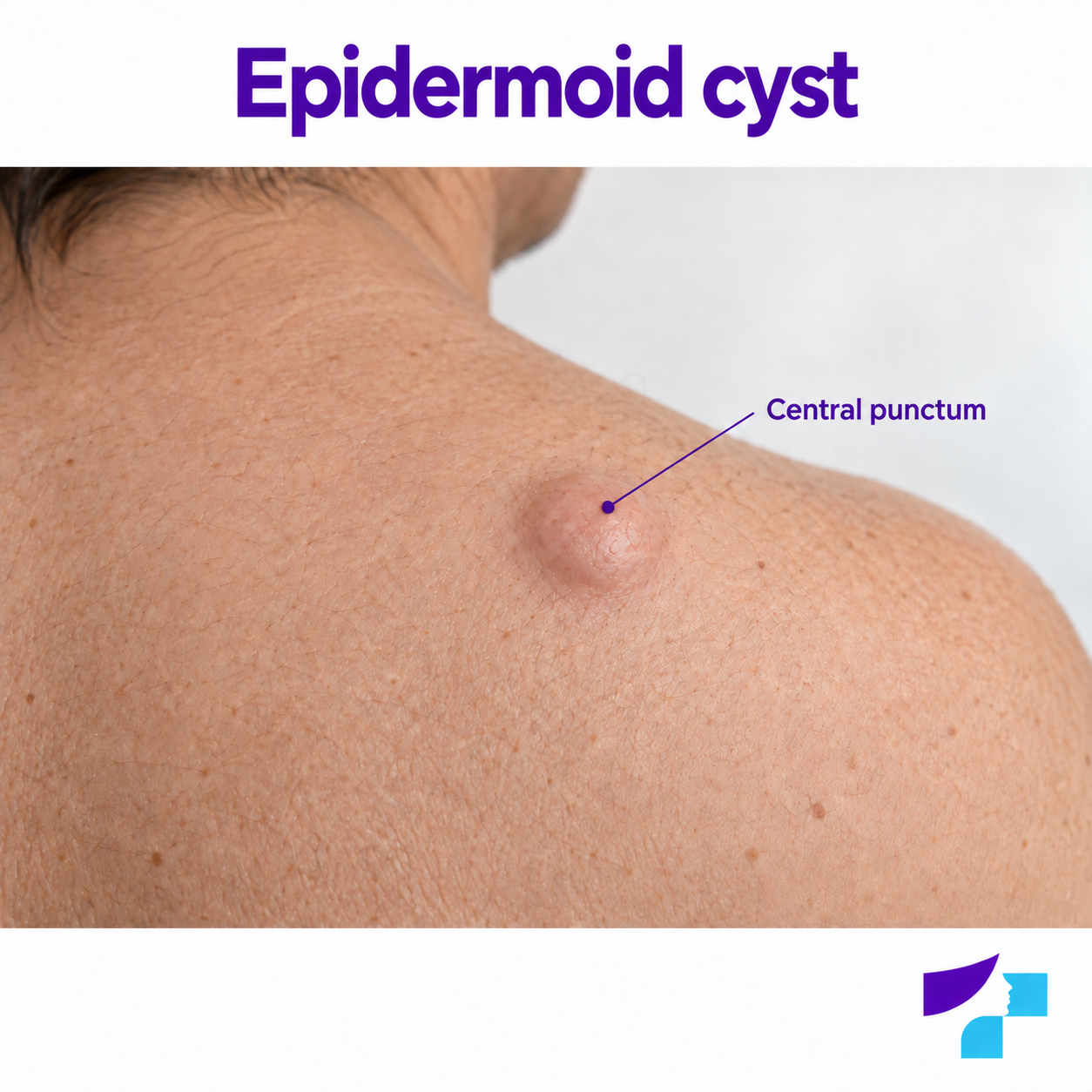
Typical features include:
- a round or dome-shaped nodule;
- slow growth;
- often measuring 1–3 cm, although it may be smaller or larger;
- a darker central point or punctum;
- mobility over deeper tissues, although the cyst may be attached to the skin;
- usually no pain unless inflammation is present;
- possible discharge of thick, white, yellowish or unpleasant-smelling material.
If a lesion grows rapidly, bleeds, ulcerates, changes colour or shape, or becomes very hard or fixed, it should not be assumed to be a simple epidermal cyst.
What is a punctum or central point?
A punctum is a small central point or opening on the surface of the cyst. It may look like a darker dot, a black point or a small pore. It is not visible on every epidermal cyst, but when present it may help the doctor identify the lesion.
The central point should not be squeezed. Squeezing may cause the cyst to rupture, become inflamed or infected.
Is a cyst with a central point always an epidermal cyst?
No. A central punctum is a typical feature of an epidermal cyst, but it is not sufficient for self-diagnosis. Other skin lesions or inflammatory conditions may look similar. A lesion that grows, hurts, changes or cannot be identified with confidence should be examined by a doctor.
What is inside an epidermal cyst?
An epidermal cyst generally contains keratin and skin-cell debris. The material may be thick, white, yellowish, cheese-like and unpleasant-smelling. This does not necessarily mean that the cyst contains pus.
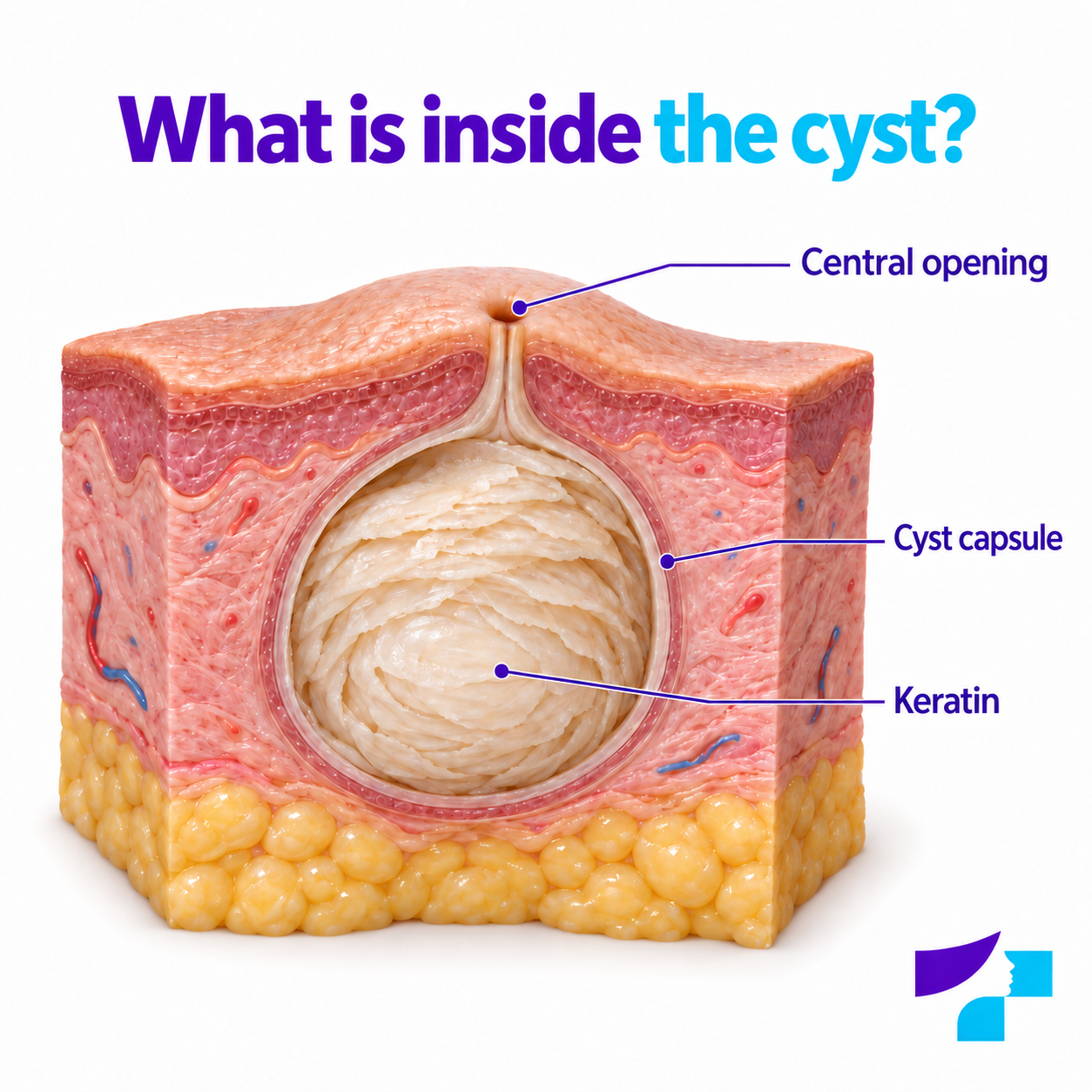
Pus is more characteristic of infection. A cyst that becomes red, hot, painful or swollen, or is associated with fever, should be examined by a doctor.
Why do epidermal cysts develop?
Epidermal cysts often develop when the upper part of a hair follicle becomes blocked or skin cells become trapped in deeper layers of the skin and continue to produce keratin. This may occur after a skin injury, scratch, surgical scar, acne or chronic skin irritation. Sometimes no clear cause can be identified.
Possible mechanisms include:
- blockage of a hair follicle;
- accumulation of keratin;
- skin cells becoming trapped in deeper layers following an injury;
- acne or inflammatory skin conditions;
- less commonly, genetic syndromes or other disorders, particularly when numerous cysts are present.
Epidermal cysts are generally not caused by poor hygiene and are not contagious.
Where do epidermal cysts most commonly develop?
Epidermal cysts may develop almost anywhere on the body, but are more commonly found:
- on the face;
- on the neck;
- on the back;
- on the chest;
- on the shoulders;
- on the scalp;
- occasionally in the genital area.
Pilar cysts, which are a different type of cyst, are also common on the scalp. A lump on the scalp is therefore not always an epidermal cyst.
Epidermal cysts on the face and small white cysts
An epidermal cyst on the face may cause cosmetic concern and can sometimes be mistaken for acne, a closed comedone or milia. Milia are very small, white keratin cysts that often develop around the eyes, on the cheeks or elsewhere on the face. They are not the same as a larger epidermal cyst with a defined capsule and deeper nodule.
Facial lesions should not be squeezed or punctured at home because scarring, changes in pigmentation and infection are particularly undesirable in this area.
Is an epidermal cyst dangerous?
Most epidermal cysts are benign and not life-threatening. They may remain for a long time without causing symptoms. A cyst may, however, become problematic if it grows, causes discomfort, repeatedly becomes inflamed, ruptures, becomes infected or causes pain.
It is also important to remember that not every lump beneath the skin is an epidermal cyst. If the diagnosis is unclear, the lesion should be assessed by a doctor.
Can an epidermal cyst be malignant?
Epidermal cysts are generally benign. Malignant change within an epidermal cyst is very rare but medically possible, and a cyst-like lesion may occasionally prove to be another diagnosis.
Medical assessment is required if the lesion:
- grows rapidly;
- becomes hard and fixed;
- bleeds or ulcerates;
- does not heal;
- changes colour, shape or surface;
- is pigmented;
- repeatedly becomes inflamed;
- is associated with fever, unexplained weight loss or pronounced fatigue.
Any new or changing pigmented skin lesion should be shown to a dermatologist.
When does an epidermal cyst become inflamed, rupture or become infected?
An epidermal cyst may become inflamed if its capsule ruptures and keratin enters the surrounding tissue. It may also become infected if bacteria enter it. One possible cause of skin infection is Staphylococcus aureus, but the exact cause of infection and need for treatment are determined by a doctor.
Signs of inflammation or infection may include:
- redness;
- swelling;
- warm or hot skin;
- pain and tenderness;
- pus or unpleasant-smelling discharge;
- rapid enlargement of the lesion;
- fever or feeling unwell.
Inflammation and infection are not the same, but they may look similar to the patient. A painful or red cyst should therefore be examined by a doctor.
What should you do if an epidermal cyst becomes red and painful?
If an epidermal cyst becomes red, hot, swollen or painful, it should not be squeezed, punctured or emptied. Until it is examined by a doctor, the skin can be kept gently clean and protected from friction or injury.
Depending on the situation, the doctor may recommend:
- observation if the inflammation is mild;
- treatment for infection when signs of bacterial infection are present;
- drainage if a substantial amount of purulent material or pressure has developed;
- planned removal later, after the inflammation has subsided.
Antibiotics may be needed for infection, but they do not remove the cyst capsule. The cyst may therefore remain or recur after the inflammation has been treated.
Why can an epidermal cyst recur after drainage?
Drainage removes the contents of the cyst, but the capsule often remains in the tissue. If the capsule remains, it can fill with keratin again and the cyst may recur.
If a cyst repeatedly becomes inflamed or returns, the doctor may recommend planned surgical removal after the inflammation has subsided.
Epidermal cyst or lipoma: how can they be distinguished?
| Feature | Epidermal cyst | Lipoma |
|---|---|---|
| Contents | Keratin and skin-cell material | Fatty tissue |
| Surface | May have a central punctum | Usually has no central punctum |
| Feel | May be firm or elastic | Often soft, rubbery and mobile |
| Inflammation | May become inflamed, rupture and discharge contents | Usually does not become inflamed |
| Unpleasant-smelling contents | May be present | Usually absent |
A reliable diagnosis cannot always be made by touch alone. If a lesion grows, hurts, lies deep or is unclear, the doctor may recommend ultrasound or removal with histological examination.
Epidermal cyst or pilar cyst: how can they be distinguished?
A pilar or trichilemmal cyst more commonly develops on the scalp and is generally associated with hair-follicle structures. It tends to be firm and smooth and often has no central punctum. An epidermal cyst is more commonly found on the face, neck, back and chest and is more likely to have a central opening.
| Feature | Epidermal cyst | Pilar cyst |
|---|---|---|
| Most common location | Face, neck, back and chest | Scalp |
| Central punctum | May often be present | Usually absent |
| Contents | Keratin | Keratin-type material |
| Number | One or several may be present | Several are often present |
| Further information | Epidermal cysts | Pilar cysts |
Epidermal cyst, boil or abscess: how can they be distinguished?
A boil or furuncle is a purulent inflammation of a hair follicle and the surrounding tissue. An abscess is a collection of pus within tissue. An inflamed epidermal cyst may resemble a boil or abscess, so self-diagnosis can be misleading.
An epidermal cyst without inflammation generally grows slowly and is painless. A boil or abscess is more likely to develop quickly, be painful, red and hot and be associated with pus or fever. Medical assessment is needed in this situation.
Epidermal cyst or acne cyst?
Deep inflammatory nodules or so-called acne cysts are not the same as epidermal cysts. Acne lesions are generally associated with comedones, inflammatory papules, pustules, nodules and a broader pattern of acne.
An epidermal cyst is more commonly a single, well-defined, encapsulated lesion containing keratin. If several inflammatory lesions are present on the face or back, acne, folliculitis and other skin conditions should also be considered.
How does a doctor diagnose an epidermal cyst?
A doctor can often diagnose an epidermal cyst by examining it. The doctor assesses the lesion’s size, shape, location, mobility, tenderness, skin colour, central punctum, signs of inflammation and whether it is attached to deeper tissues.
The doctor may ask:
- how long the cyst has been present;
- whether it is growing;
- whether it hurts or becomes inflamed;
- whether there has been a previous injury;
- whether any material has discharged from it;
- whether similar lesions have occurred before;
- whether family members have similar cysts.
When are ultrasound, dermoscopy or histology needed?
A typical epidermal cyst can often be recognised clinically. Further tests may be needed if the diagnosis is unclear or the lesion is deep, atypical, growing rapidly or must be distinguished from another lesion.
Dermoscopy helps assess structures on the skin surface, particularly when a lesion is pigmented or needs to be distinguished from another skin lesion.
Soft-tissue ultrasound can help determine whether a lesion is cystic or solid, how deep it lies and whether it is connected to surrounding tissue.
Histological examination means examining the removed lesion under a microscope. It is particularly important if the diagnosis is not completely clear, the lesion is atypical or recurrent, or another process needs to be excluded.
How are epidermal cysts treated?
Treatment depends on the size and location of the cyst, its symptoms and whether it is inflamed.
Possible approaches include:
- observation if the cyst is small, painless and unchanged;
- treatment of inflammation or infection;
- drainage if the cyst is very painful, tense or purulent;
- surgical removal together with the capsule if the cyst grows, causes discomfort, repeatedly becomes inflamed or the diagnosis is unclear;
- histological examination after removal when medically indicated.
More complete removal of the cyst together with its capsule reduces the risk of recurrence. If only the contents are emptied, the capsule may remain and the cyst may fill again.
When should an epidermal cyst be removed?
Removal of an epidermal cyst may be recommended if it:
- grows;
- hurts;
- repeatedly becomes inflamed;
- discharges contents;
- catches on clothing or interferes with shaving or daily activities;
- is in a visible location and causes significant cosmetic concern;
- recurs after drainage;
- cannot be diagnosed with confidence.
An epidermal cyst is often easier to remove electively while it is not inflamed. During inflammation, the capsule may be ruptured or firmly attached to surrounding tissue, making complete removal more difficult.
The procedure, anaesthesia, stitches, healing, scarring and suture removal are described in more detail in the article “Cyst removal”.
What should not be done at home?
An epidermal cyst should not be squeezed, punctured with a needle, cut, burned, scratched or emptied at home. Caustic substances, alcohol, lemon, baking soda, toothpaste, apple cider vinegar and other aggressive products should not be applied to it.
These actions can cause rupture of the cyst, spread inflammation, infection, scarring, changes in pigmentation and recurrence. They may also delay an accurate diagnosis if the lesion is not actually an epidermal cyst.
Epidermal cysts in specific patient groups
Children
A new or growing skin lesion in a child should be assessed carefully because not every lump beneath the skin is a simple epidermal cyst. Some lesions may be congenital and require specific diagnostic evaluation.
During pregnancy
Pregnant patients should not use medicines or aggressive topical products without medical advice. A doctor should be consulted if a cyst hurts, becomes inflamed or is located in the genital or breast area.
People with weakened immunity
Infections may be more severe in people with diabetes, immunosuppression, cancer treatment or another cause of weakened immunity. In this situation, a painful, red or purulent cyst should be assessed without delay.
Common myths about epidermal cysts
| Myth | Fact |
|---|---|
| An epidermal cyst is a sebaceous cyst | This is often an inaccurate term; the cyst usually contains keratin. |
| Atheroma is always an accurate diagnosis | The word is used broadly in everyday language, but the diagnosis should be confirmed by a doctor. |
| An epidermal cyst is filled with fat | It is generally filled with keratin and skin-cell material. |
| White contents are always pus | The material is often keratin rather than pus. |
| An epidermal cyst is contagious | An ordinary epidermal cyst is not contagious. |
| It is safe to squeeze a cyst | Squeezing may cause inflammation, infection and scarring. |
| An epidermal cyst must always be removed | Some small, painless and unchanged cysts can be monitored. |
| Antibiotics cure an epidermal cyst | They may help treat a bacterial infection but do not remove the capsule. |
| A cyst with a central point is always an epidermal cyst | A central punctum is characteristic, but the diagnosis is made by a doctor. |
| A cyst cannot return after drainage | Drainage often leaves the capsule in place, so recurrence is possible. |
| An epidermal cyst develops because of poor hygiene | Cyst formation is generally not associated with inadequate hygiene. |
Summary
An epidermal or epidermoid cyst is a common and usually benign skin lesion that is generally filled with keratin rather than fat. It may look like a round lump beneath the skin, sometimes with a central punctum. Although most epidermal cysts are not dangerous, they may become inflamed, rupture, become infected or be mistaken for another lesion.
A cyst should not be squeezed or punctured at home. A lesion that grows, hurts, becomes red, bleeds, ulcerates, changes or cannot be identified with confidence requires medical assessment.
Important: this information is intended for educational purposes and does not replace a dermatologist consultation, diagnosis or treatment. Do not use prescription medicines, antibiotics, corticosteroids, caustic substances or invasive methods at home without medical advice. Children, pregnant patients, people with weakened immunity and anyone with a lesion on the face, head, neck, genital or breast area should consult an appropriate doctor.
References and authoritative sources
- DermNet: causes, features, complications and treatment of epidermoid cysts
- DermNet: types of cutaneous cysts and pseudocysts
- UK National Health Service (NHS): features and treatment of skin cysts
- Cleveland Clinic: features, causes, diagnosis and treatment of epidermal inclusion cysts
- Mayo Clinic: symptoms and causes of epidermoid cysts
- US National Center for Biotechnology Information medical library: diagnosis and treatment of epidermal inclusion cysts
- American Academy of Family Physicians: minimal excision technique for epidermoid cysts
- Merck Manual: principles of diagnosis and treatment for cutaneous cysts
