A pilar cyst, also called a trichilemmal cyst, is a usually benign, slow-growing nodule in or beneath the skin that develops from hair follicle structures. It most commonly occurs on the scalp, so people often notice it while washing, brushing or touching their hair.
Important: a pilar cyst on the scalp is not a “brain cyst”, an arachnoid cyst or a lesion inside the skull. It is a skin or subcutaneous cyst beneath the hair. Searching for a “cyst in the head” can cause confusion, so it is important to distinguish a lump that can be felt on the scalp from a cyst found on brain imaging.
Most pilar cysts are not dangerous and cause no symptoms for a long time. However, not every scalp lump is a pilar cyst. A lesion should be assessed by a doctor if it grows rapidly, becomes painful, red, hot, hard or fixed, bleeds, ulcerates or cannot be clearly identified.
What is a pilar or trichilemmal cyst?
Pilar cyst and trichilemmal cyst mean the same thing. “Trichilemmal” refers to its origin in the outer root sheath of a hair follicle, while the shorter term “pilar cyst” is more commonly used in everyday language.
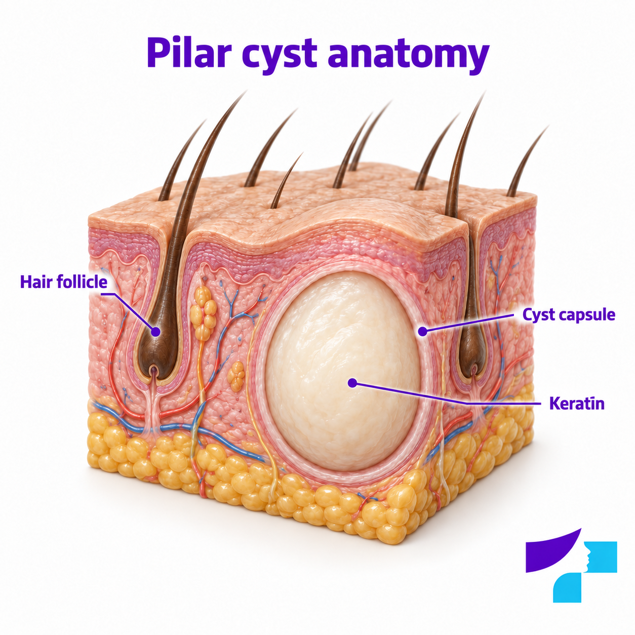
It is a skin or subcutaneous cyst, not a cyst of an internal organ. It develops in the scalp skin or subcutaneous tissue, rather than in the brain, inside the skull or in other internal tissues. The cyst wall, or capsule, encloses keratin: dense protein material associated with skin, hair and nail cells.
In simple terms, a pilar cyst is a small, encapsulated “sac” beneath the skin, filled with keratin, that most commonly develops in areas with many hair follicles.
Why do pilar cysts most commonly develop on the scalp?
The scalp contains a very large number of hair follicles. A hair follicle is the structure in the skin from which a hair grows. It has several parts, including the outer root sheath: a layer of cells involved in forming the structures that support hair growth.
A pilar cyst develops when cells and keratin begin to accumulate within an enclosed capsule in this area. This is why pilar cysts are most commonly found on the scalp, although they can less frequently occur on other hair-bearing parts of the body.
The term “trichilemmal keratinization” means that the cyst contents form through a process similar to the conversion of outer root sheath cells into keratin material. The key point for patients is that the capsule contains dense keratin, rather than dirt, pus or fat.
What does a pilar cyst look and feel like?
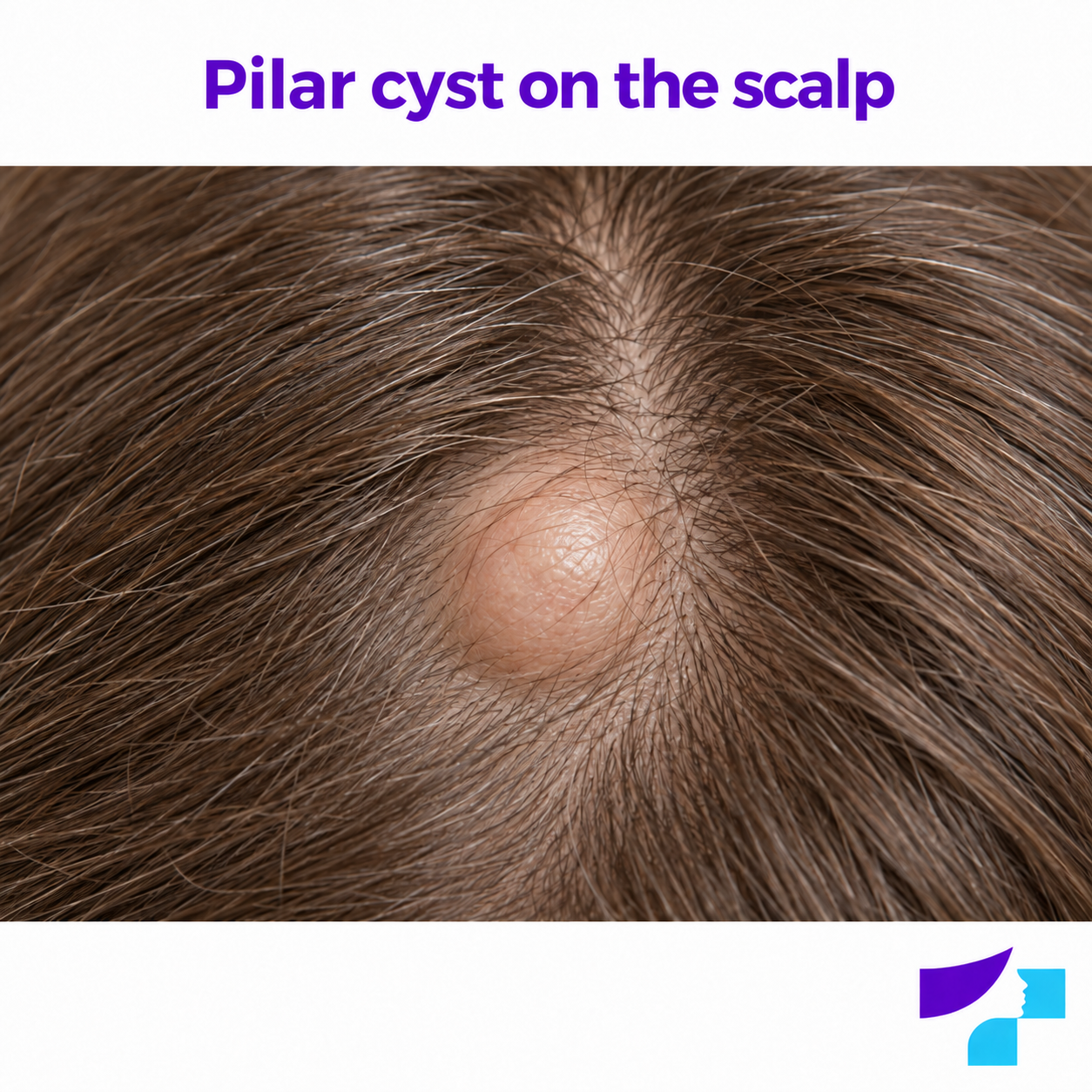
A pilar cyst on the scalp is usually a smooth, round or oval, firm, mobile nodule beneath the skin. The overlying skin often looks normal, and the cyst is usually painless unless it becomes inflamed, ruptures or becomes infected.
| Feature | Typical of a pilar cyst |
|---|---|
| Shape | Round or oval |
| Surface | Smooth |
| Consistency | Firm, with some elasticity |
| Mobility | Usually mobile beneath the skin |
| Skin colour | Often unchanged |
| Pain | Usually painless unless inflamed |
| Growth | Slow, over months or years |
| Central punctum | Usually no obvious opening |
People often notice a pilar cyst by touch rather than in a mirror: while washing, brushing or drying their hair, or accidentally touching their scalp. Patients may describe it as a “hard lump under the hair”, a “scalp lump”, a “cyst under the skin on the head” or a “hard bump beneath the scalp”.
Small cysts may be pea-sized, but some enlarge over time and interfere with brushing, hairstyling, wearing headwear or sleeping on the affected area.
Can there be more than one pilar cyst?
Yes, pilar cysts can be multiple. Some people have a single scalp nodule, while others have several similar nodules in different parts of the scalp.
Multiple pilar cysts may suggest a familial tendency, especially if parents, siblings or other close relatives have had similar lesions. Multiple cysts do not in themselves indicate cancer, but any new, growing or different-looking lesion should be assessed by a doctor.
Are pilar cysts inherited?
Pilar cysts can be sporadic, meaning that they develop without a clear family history. However, some people have a familial predisposition. Medical literature describes cases of autosomal dominant inheritance, meaning that the tendency can be passed down through generations.
If several family members have had scalp cysts, this may explain why you develop them too. It does not mean that every cyst will need removal, but it does mean that their number, growth and changes deserve attention.
Are pilar cysts more common in women?
Some medical sources report that pilar cysts are more often seen in women and adults. However, they can also occur in men, younger people and, less commonly, children.
Sex and age alone are not sufficient for diagnosis. More important factors include the appearance and growth of the lesion, pain, inflammation, mobility, similar lesions in the family and any signs suggesting another diagnosis.
Pilar cyst or epidermal cyst: how can they be distinguished?
Pilar and epidermal cysts are similar because both are skin or subcutaneous cysts containing keratin. However, they are not the same.
| Feature | Pilar cyst | Epidermal cyst |
|---|---|---|
| Most common location | Scalp | Face, neck, back, chest and other areas |
| Origin | Outer root sheath of a hair follicle | Epidermal cells or cells of the upper part of a follicle |
| Central punctum | Usually absent | A punctum may often be present |
| Contents | More compact keratin | Thick keratin material, sometimes with an unpleasant odour |
| Number of cysts | Often multiple | Often single, but may be multiple |
| Familial tendency | Possible | Generally less characteristic |
| Definitive confirmation | Histology after removal | Histology after removal |
In practice, pilar and epidermal cysts cannot always be distinguished with certainty by appearance alone. Both can be smooth subcutaneous nodules, and histological examination after removal often provides definitive confirmation.
For a detailed explanation of epidermal cysts, see Epidermal cysts.
Is a pilar cyst an atheroma or a sebaceous cyst?
In everyday language, people often call subcutaneous cysts “atheromas”, “fatty cysts” or “sebaceous cysts”. These terms can be inaccurate. Many so-called sebaceous cysts do not arise from sebaceous glands, but are epidermal or pilar cysts containing keratin.
A doctor may suspect a pilar cyst if a scalp lump is firm, smooth, slow-growing and has no visible central punctum. A central punctum and a location elsewhere on the body may favour an epidermal cyst. However, clinical examination and, if the lesion is removed, histology establish the most accurate diagnosis.
Pilar cyst or arachnoid cyst: why must they not be confused?
An arachnoid cyst is a cavity filled with cerebrospinal fluid in the region of the membranes surrounding the brain or spinal cord. It is diagnosed using imaging, such as magnetic resonance imaging or computed tomography, and is managed by neurology or neurosurgery specialists.
A pilar cyst is a completely different diagnosis. It is a palpable nodule in or beneath the scalp skin that develops from hair follicle structures. It is not a brain cyst and does not usually cause neurological symptoms.
If a “cyst in the head” was found on an MRI or CT scan, it is outside the scope of this article. If a lump can be felt on the scalp beneath the hair, a pilar cyst is one possible explanation.
Pilar cyst, lipoma, lymph node or abscess: when should you be concerned?
Not every scalp lump is a cyst. A subcutaneous lesion may also be a lipoma, an enlarged lymph node, an abscess, a boil, a dermoid cyst, a pilomatricoma or, less commonly, a suspicious skin tumour.
| Lesion | Typical features |
|---|---|
| Pilar cyst | Smooth, firm, mobile scalp nodule, usually without a central opening |
| Lipoma | Softer, rubbery, slow-growing fatty tissue lump |
| Lymph node | Usually in typical lymph node locations; may be associated with infection |
| Abscess | Painful, red, hot and swollen; pus may be present |
| Boil | Inflamed, painful skin lesion around a hair follicle |
| Dermoid cyst | May be congenital; more common in certain areas of the head and face |
| Pilomatricoma | A harder lesion, more common in children and young people; may calcify |
| Skin tumour | May grow, ulcerate, bleed, change its surface or become fixed |
Particularly important: any new or changing pigmented scalp lesion should be assessed by a dermatologist. Pigmented scalp lesions are difficult to assess yourself because they are covered by hair.
For a broader overview of skin and subcutaneous cyst types, see Skin and subcutaneous cysts.
Is a pilar cyst dangerous?
An ordinary pilar cyst is usually not dangerous. It is not contagious, is not associated with poor hygiene and is generally not life-threatening. Some pilar cysts remain stable for years without causing medical problems.
However, a pilar cyst may become troublesome if it:
- enlarges;
- is repeatedly injured by a comb, razor or headwear;
- causes pain or pressure;
- becomes inflamed;
- ruptures;
- becomes infected;
- interferes with everyday care;
- causes cosmetic or psychological distress;
- looks atypical or changes rapidly.
The key is to assess whether the lesion behaves like a typical, uncomplicated pilar cyst, rather than assuming that it is definitely harmless.
Can a pilar cyst be malignant?
An ordinary pilar cyst is almost always benign. Malignant transformation is very rare. However, a rare lesion known as a proliferating trichilemmal tumour has been described; it can develop from trichilemmal cyst-type structures or resemble a pilar cyst.
Particular attention should therefore be paid to cysts that:
- grow rapidly;
- become large within a short time;
- change shape;
- ulcerate or bleed;
- become hard and fixed;
- repeatedly become inflamed;
- return after removal;
- look different from the other cysts;
- cause persistent pain or pressure.
In these cases, a doctor may recommend removal and histological examination to assess the tissue under a microscope.
What is a proliferating trichilemmal tumour?
A proliferating trichilemmal tumour is a rare skin lesion with histological features related to trichilemmal or pilar structures. It is not the same as an ordinary pilar cyst.
A typical pilar cyst grows slowly and is a well-defined, uncomplicated nodule. A proliferating trichilemmal tumour may grow faster, become larger and sometimes ulcerate or look atypical. Most of these tumours are benign or locally confined, although malignant forms have also been described rarely.
Patients are not expected to distinguish these diagnoses themselves. The practical rule is simple: a rapidly growing, atypical, ulcerated, bleeding or repeatedly inflamed scalp lump should be assessed by a doctor.
When can a pilar cyst become inflamed, rupture or hurt?
A pilar cyst without inflammation is usually painless. Pain is more likely when the cyst:
- is injured;
- is squeezed or punctured;
- ruptures beneath the skin;
- becomes inflamed;
- becomes infected;
- is in an area exposed to repeated pressure;
- has become large and presses on surrounding tissues.
If the capsule ruptures, keratin may enter the surrounding tissues and trigger an inflammatory reaction. The lesion can then become painful, tender, swollen and red. If infection develops, pus, warmth, severe pain or fever may occur.
What should you do if a scalp cyst becomes red or painful?
If a scalp lump becomes red, painful, hot or swollen, do not squeeze, puncture, cut or try to empty it at home.
Safe steps:
- Avoid pressing on or irritating the lesion.
- Do not apply caustic or harsh products.
- Keep the area clean and dry.
- If the cyst opens by itself, cover it with a clean dressing.
- Contact a family doctor, dermatologist or surgeon.
- Seek help more urgently if there is fever, pus, rapidly increasing swelling or a feeling of being unwell.
An inflamed or infected cyst sometimes requires drainage or medication. However, drainage is not the same as complete cyst removal: if the capsule remains in the skin, the cyst can recur.
Can a pilar cyst affect the hair?
A small, uncomplicated pilar cyst does not usually cause hair loss. A larger cyst can raise the skin, interfere with brushing or cause local discomfort. If it becomes inflamed, ruptures or is injured, the surrounding skin may become sensitive, and hair in that area may appear thinner because of inflammation, pressure or treatment.
A small scar may remain after removal. Hair may grow differently or fail to regrow fully within the scar, depending on the lesion's size and location, inflammation, the size of the incision and individual healing. For details of the procedure, see Cyst removal.
How does a doctor diagnose a pilar cyst?
A doctor can often recognise a pilar cyst by examining and feeling it. The assessment considers:
- where the lesion is located;
- its size;
- whether it is mobile;
- whether it is painful;
- whether there is a central opening;
- whether the overlying skin has changed;
- whether inflammation is present;
- whether there are multiple cysts;
- whether relatives have had similar lesions;
- how quickly the lesion is growing.
If the findings are typical — a smooth, firm, slow-growing, mobile scalp nodule without signs of inflammation — additional investigations are not always necessary.
When are ultrasound, biopsy or histology needed?
Additional investigations may be needed if the diagnosis is unclear or atypical features are present.
| Investigation | When it may be needed |
|---|---|
| Ultrasound | If the lesion is deeper, unclear, hard or fixed, or needs to be distinguished from a lipoma, lymph node, abscess or another lesion |
| Dermoscopy | To assess the skin surface, pigmentation or a suspicious skin lesion |
| Biopsy | If an atypical or tumour-like process is suspected |
| Histology | After removal, to confirm the diagnosis microscopically |
| CT or MRI | Rarely, if the lesion is very deep, atypical or associated with anatomically complex areas of the head and neck |
A removed cyst is often sent for histological examination, especially if it was rapidly growing, atypical, recurrent, inflamed or clinically uncertain.
How are pilar cysts treated?
Treatment depends on symptoms, size, inflammation and the patient's needs.
A small, painless, slow-growing cyst that causes no problems can be monitored. If it enlarges, interferes with daily activities, is injured or causes discomfort, surgical removal may be considered.
If the cyst is inflamed or infected, a doctor may first treat the inflammation. Drainage is sometimes needed, particularly if pus has accumulated. Antibiotics may be required when there are signs of bacterial infection, but antibiotics alone do not usually remove the capsule and therefore do not eliminate the cyst itself.
Definitive treatment of a troublesome or recurrent cyst usually requires removal of the cyst together with its capsule.
When should a pilar cyst be removed?
A pilar cyst does not need removal simply because it exists. Removal is generally considered if the cyst:
- grows;
- becomes painful;
- repeatedly becomes inflamed;
- ruptures;
- interferes with brushing or washing the hair;
- is repeatedly injured;
- causes significant cosmetic distress;
- is large;
- is atypical;
- cannot be clearly diagnosed;
- requires histological examination;
- returns after previous treatment.
Removal is often easier when the cyst is not inflamed. During inflammation, tissues are more sensitive, complete capsule removal may be harder and healing may be more complicated. If a cyst repeatedly causes problems, it is better to discuss planned removal with a doctor in good time rather than wait for another episode of inflammation.
Does the hair need to be shaved before pilar cyst removal?
Not always. This depends on the cyst's size and location, hair thickness, the surgical approach and the doctor's technique. In some cases, only a small area needs to be exposed; in others, a little hair may need to be shaved around the incision.
For more information about anaesthesia, stitches, healing, scarring, hair shaving and aftercare, see Cyst removal.
Can a pilar cyst return after removal?
Yes, a pilar cyst can recur. The risk is greater if the capsule was not completely removed, if the cyst had previously ruptured or become inflamed, or if the person is predisposed to developing multiple pilar cysts.
Sometimes a new cyst develops over time in the same or another area of the scalp after one has been removed. This does not necessarily mean that the previous procedure was performed incorrectly: people with a familial tendency may develop new nodules.
What should not be done at home?
Do not squeeze, puncture, cut, scratch, burn or try to empty a pilar cyst yourself. This can cause:
- rupture beneath the skin;
- inflammation;
- bacterial infection;
- pus formation;
- pain and swelling;
- scarring;
- more difficult removal later;
- delayed diagnosis if the lesion is not a cyst.
There is no evidence that alcohol compresses, lemon, baking soda, toothpaste, apple cider vinegar, caustic products or “drawing” ointments safely eliminate pilar cysts. These methods can burn or irritate the scalp and worsen the condition.
When should you definitely see a doctor?
See a doctor if a pilar cyst or a similar scalp lump:
- grows rapidly;
- becomes hard and fixed;
- becomes painful, hot, red or swollen;
- discharges pus, blood or unpleasant fluid;
- repeatedly becomes inflamed;
- interferes with brushing or washing the hair;
- is repeatedly injured;
- is accompanied by fever or feeling unwell;
- appears in a child, pregnant woman, older adult or person with a weakened immune system;
- changes shape, surface or colour;
- bleeds, ulcerates or fails to heal;
- enlarges rapidly after a long stable period;
- cannot be confidently identified as a pilar cyst.
Any new or changing pigmented scalp lesion should be assessed by a dermatologist.
Pilar cysts in children, pregnant women and people with weakened immunity
A scalp lump in a child requires particular care because possible diagnoses include congenital lesions, dermoid cysts, pilomatricoma and other skin or subcutaneous lesions.
Treatment during pregnancy is individualised. Removal may be postponed if the cyst is uncomplicated and causes no problems. If it hurts, becomes inflamed, grows rapidly or becomes infected, medical assessment is needed.
Inflammation and infection may be more severe in people with weakened immunity, diabetes or chronic diseases. A painful, red or pus-producing lesion should be assessed promptly in these circumstances.
Common myths about pilar cysts
| Myth | Fact |
|---|---|
| Pilar cyst and trichilemmal cyst are different diseases | They are names for the same lesion |
| A pilar cyst is the same as an epidermal cyst | They are similar but distinct types of cyst |
| A pilar cyst is the same as an arachnoid cyst | No; an arachnoid cyst is associated with the membranes surrounding the brain or spinal cord |
| A scalp lump is always a pilar cyst | It may also be a lipoma, lymph node, abscess, pilomatricoma or another lesion |
| A pilar cyst is contagious | It is not contagious |
| Pilar cysts result from poor hygiene | Usually not; hair follicle structures and genetic predisposition are relevant |
| It is safe to squeeze a pilar cyst | Squeezing can cause inflammation and infection |
| Antibiotics cure a pilar cyst | Antibiotics can treat infection but do not remove the capsule |
| A pilar cyst must always be removed | Not always; uncomplicated cysts can be monitored |
| A pilar cyst always disappears by itself | The capsule does not usually disappear on its own |
| A pilar cyst always causes hair loss | Usually it does not |
| The hair must always be shaved before removal | This depends on the circumstances |
| A cyst never returns after removal | Recurrence or new cysts are possible |
Summary
A pilar or trichilemmal cyst is a benign skin or subcutaneous cyst that most commonly develops on the scalp and is associated with the outer root sheath of a hair follicle. It is usually smooth, firm, round, mobile and painless. Unlike an epidermal cyst, it generally has no obvious central punctum.
A pilar cyst is neither an arachnoid cyst nor a brain cyst. It is a palpable subcutaneous lesion beneath the hair. Although most pilar cysts are not dangerous, they may become inflamed, rupture, become infected, grow or interfere with daily life. A rapidly growing, painful, bleeding, ulcerated, hard, fixed or unexplained scalp lesion should be assessed by a doctor.
The safest way to treat a troublesome pilar cyst is removal following medical assessment, rather than squeezing or emptying it yourself at home.
Medical disclaimer
This information is educational and does not replace a dermatologist consultation, diagnosis or treatment. Any new, growing, painful, bleeding, ulcerated, rapidly changing, inflamed, infected, pigmented, hard, fixed or unexplained skin or subcutaneous lesion, including on the scalp, should be assessed by a doctor. Do not use prescription medicines, antibiotics, corticosteroids, caustic products or invasive home procedures without medical guidance. Children, pregnant women, people with weakened immunity and anyone with lesions on the face, head, neck or intimate areas should consult an appropriate doctor.
References and authoritative sources
- Cleveland Clinic: causes, signs and removal of pilar or trichilemmal cysts
- US National Center for Biotechnology Information medical library (NCBI Bookshelf), StatPearls: diagnosis and treatment of pilar cysts
- US National Center for Biotechnology Information medical library (NCBI Bookshelf), StatPearls: histopathological evaluation of skin cysts
- DermNet: features, origin and treatment of pilar or trichilemmal cysts
- DermNet: proliferating trichilemmal lesions and their treatment
- DermNet: types of cutaneous cysts and pseudocysts
- DermNet: features of epidermoid cysts and differences from pilar cysts
- UK National Health Service (NHS): signs and treatment of skin cysts
