Angiomas are benign vascular skin growths that most often appear as red, dark red, purple or bluish dots, spots or small bumps. People often call them “red moles”, but medically an angioma is not the same as a pigmented mole.
The most common type of angioma in adults is a cherry angioma. It is usually harmless and does not always require treatment. However, not every red skin growth is an angioma. If a lesion grows quickly, changes, bleeds, hurts, itches or looks different from other skin lesions, it should be shown to a dermatologist.
In brief: the most important facts about angiomas
- Angiomas are usually benign vascular growths on the skin.
- Cherry angiomas are more common in adults, especially after the age of 30-40.
- They may be flat or raised, bright red, dark red, purple or bluish.
- A typical angioma is usually not cancer and does not turn into cancer.
- Angiomas do not always need to be removed if they do not cause discomfort and the diagnosis is clear.
- Angioma removal may be appropriate if the lesion bleeds, is repeatedly irritated or causes cosmetic concern.
- Removal methods include laser treatment, electrocoagulation, cryotherapy or surgical removal.
- An angioma should not be cut, burned or treated with caustic products at home.
- Accurate diagnosis may require dermoscopy.
- If the diagnosis is unclear, the removed lesion may be sent for histological examination.
When should an angioma be shown to a dermatologist?
You should see a dermatologist if:
- the lesion grows quickly;
- it bleeds often or bleeds without clear trauma;
- its colour, shape or size changes;
- the lesion hurts, itches, ulcerates or becomes inflamed;
- it looks different from other skin lesions;
- many new red dots appear on the skin in a short time;
- it is unclear whether the lesion really is an angioma;
- the lesion is in an area where it is regularly irritated by clothing, shaving or jewellery;
- the patient has an increased risk of skin cancer or has previously had a skin tumour.
A red skin lesion is not always a harmless angioma. Self-diagnosis based on internet images is therefore not safe. In such cases, a professional skin lesion examination is important.
What is an angioma?
An angioma is a benign skin growth related to blood vessels or the proliferation of cells in the vessel wall. It may be superficial and bright red, or deeper and bluish-purple. Some angiomas are level with the skin, while others are slightly raised.
Patients often notice an angioma as a small red dot on the skin. It may appear on the chest, abdomen, back, shoulders, arms, face or other areas of the body. Some people have one angioma, while others develop many angiomas over time.
A typical cherry angioma is usually benign. However, other skin lesions may look similar, including pyogenic granuloma, an inflamed mole, basal cell carcinoma or, rarely, amelanotic melanoma. This is why a dermatologist’s assessment is important when a lesion does not look typical.
Common types of angiomas and similar vascular skin lesions
| Type | What it looks like | Where it most often appears | Is it usually dangerous? | When should it be checked? |
|---|---|---|---|---|
| Cherry angioma | A small red, dark red or purple dot or bump | Trunk, chest, abdomen, back, arms | Usually not dangerous | If it bleeds, grows, changes or causes discomfort |
| Spider angioma | A red central point with fine blood vessels radiating around it | Face, neck, upper chest | Often benign | If there are many lesions or other symptoms are present |
| Venous lake | A soft bluish or purple bump | Lips, ears, face | Usually benign | If it changes, bleeds or needs to be distinguished from another lesion |
| Angiokeratoma | A dark red, purple or almost black, sometimes rough lesion | Legs, trunk, genital area | Often benign | If it bleeds, hurts, appears in large numbers or the diagnosis is unclear |
| Infantile haemangioma | A red or bluish vascular growth in a child | Head, neck, trunk | Some shrink on their own | If it grows quickly, ulcerates or is located in a high-risk area |
| Vascular malformation | A patch or deeper vascular change, often present from birth | Depends on the type | Not the same as a cherry angioma | Requires individual specialist evaluation |
Cherry angioma
Cherry angioma is the most common angioma in adults. It is usually a small, round, clearly defined red or dark red growth. Its size is often 1 to 5 mm, although sometimes it may be larger. At first it may be flat, later becoming dome-shaped or slightly raised.
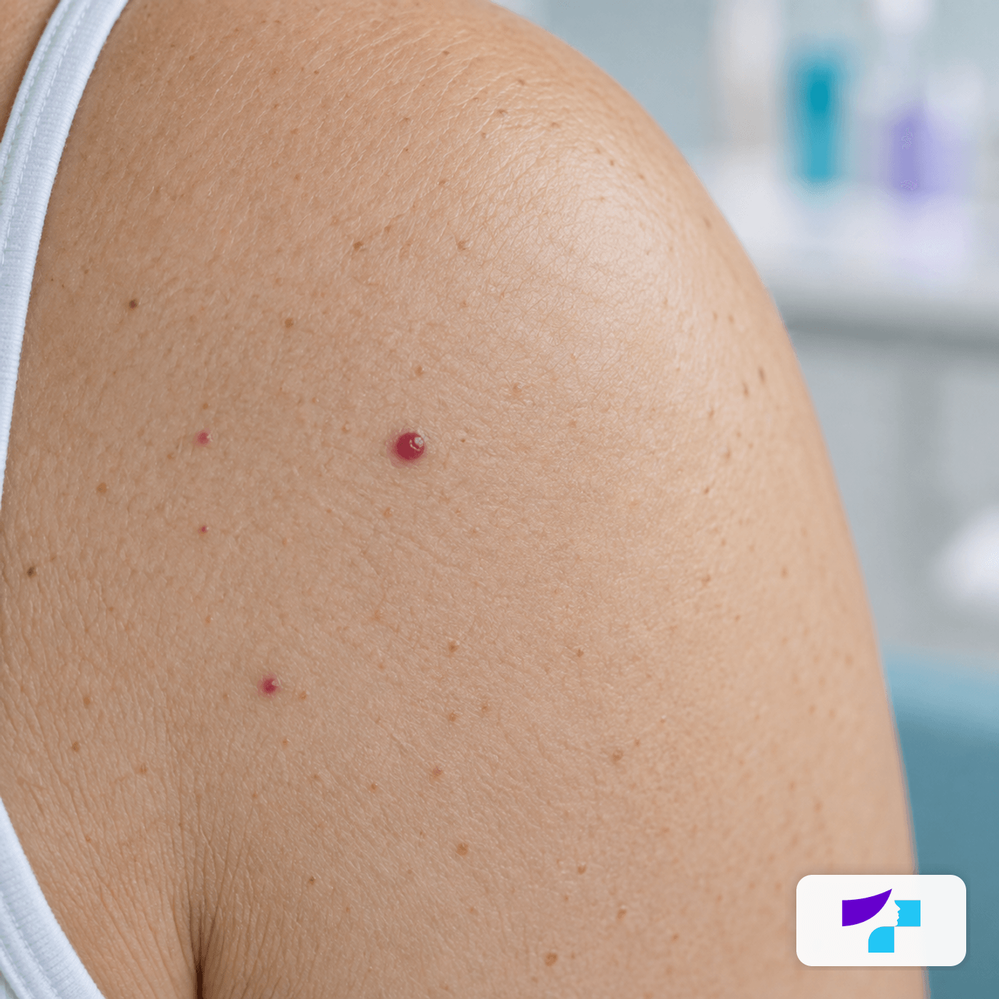
The visual shows a typical cherry angioma as a small red vascular growth on adult skin.
Cherry angiomas most often appear on the trunk: the back, abdomen, chest, shoulders and arms. Their number may increase with age. This in itself is usually not dangerous.
The exact cause of cherry angiomas is often unknown. Age, genetic predisposition, hormonal changes and individual vascular characteristics of the skin may play a role. They are not contagious and are not caused by an infection.
Why do angiomas form?
The formation of angiomas is not always linked to one specific cause. Possible factors include:
- age;
- genetic predisposition;
- individual vascular changes;
- hormonal changes, including pregnancy;
- skin ageing;
- the influence of certain medicines or external factors;
- in the case of spider angiomas - also the context of liver disease or hormonal factors.
One or a few cherry angiomas usually do not indicate a serious disease. If many new angiomas appear suddenly within a short period, especially together with fatigue, jaundice, unexplained weight loss, a tendency to bleed or other symptoms, medical advice should be sought.
What does an angioma look like?
A typical angioma may be:
- bright red, dark red, purple or bluish;
- flat or raised;
- smooth and clearly defined;
- very small or several millimetres in size;
- painless and not itchy;
- prone to bleeding if traumatised.
If a lesion becomes painful, ulcerates, bleeds without reason, grows rapidly or changes colour, it should no longer be treated as a simple cosmetic issue.
Angioma, mole, haemangioma or melanoma: what is the difference?
| Lesion | Typical colour | Related to blood vessels? | What is important for the patient? |
|---|---|---|---|
| Angioma | Red, purple or bluish | Yes | Usually benign, but should be checked if it changes or bleeds |
| Cherry angioma | Bright red to dark purple | Yes | Common in adults and usually harmless |
| Pigmented mole | Light brown, brown or dark | Usually no | Changes in colour, shape, borders and size should be monitored |
| Haemangioma in a child | Red or bluish | Yes | In children, assessment depends on age, location and growth |
| Pyogenic granuloma | Red, fast-growing, bleeds easily | Yes | Often requires medical assessment and sometimes removal |
| Melanoma or another skin tumour | May be brown, black, pink, red or non-pigmented | Not always | A changing or atypical lesion should be checked promptly |
How does a dermatologist diagnose an angioma?
Diagnosis begins with a skin examination. The dermatologist evaluates the lesion’s colour, shape, borders, surface, size and location. Dermoscopy is often used - an examination of the skin lesion with special magnification and lighting.
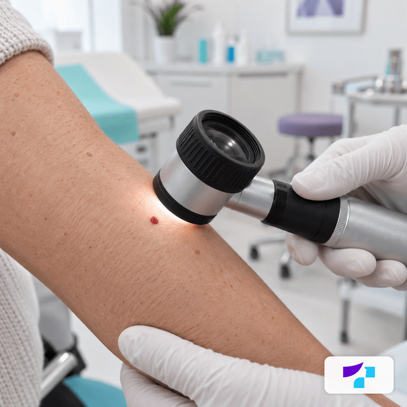
The visual shows a dermatologist examining a small red skin lesion with a dermoscope to assess its features and distinguish an angioma from other lesions.
The doctor may ask:
- when the lesion appeared;
- whether it has changed;
- whether it bleeds;
- whether it is being irritated or traumatised;
- whether there is pain, itching or inflammation;
- whether any other new lesions have appeared recently;
- whether the patient has had atypical moles or skin tumours before.
If the diagnosis is unclear, the lesion may be removed and sent for histological examination. Histology helps confirm the diagnosis in the laboratory.
Are angiomas dangerous?
Most cherry angiomas are not dangerous. They are benign, common and usually do not affect general health. They may cause cosmetic concern or bleed if traumatised.
The most important risk is mistaking an angioma for another skin lesion. If a “red angioma” looks atypical, grows quickly, bleeds without trauma or ulcerates, it should be checked by a dermatologist.
Do angiomas need treatment?
Angioma treatment is not always necessary. If the diagnosis is clear, the lesion does not cause discomfort and does not change, it may simply be observed.
Angioma removal may be appropriate if:
- the angioma bleeds regularly;
- it is irritated by clothing, shaving or jewellery;
- it is located in a cosmetically bothersome area;
- it causes discomfort to the patient;
- the diagnosis is not fully clear and histology is needed.
Angioma removal methods
| Situation | Possible method | What can be achieved | Important caution |
|---|---|---|---|
| Small, typical cherry angioma | Laser or electrocoagulation | Reduce or remove the red lesion | The diagnosis should be confirmed first |
| Raised angioma that rubs or bleeds | Electrocoagulation, shave excision or surgical removal | Remove a bothersome lesion | Scabbing, pigmentation changes or a scar are possible |
| Atypical or unclear lesion | Surgical removal with histology | Clarify the exact diagnosis | Laser is not the best choice if histology is needed |
| Multiple small vascular lesions | Laser treatment after assessment | Improve the cosmetic appearance of the skin | Skin phototype and sun exposure must be considered |
| Bluish or deeper changes | Individual treatment plan | Depends on the type of lesion | Additional diagnostics may be needed |
Laser treatment for angioma removal
Laser removal of angiomas is often suitable for small, superficial vascular lesions. A vascular laser targets haemoglobin within the blood vessels and helps reduce or remove the red lesion. More about the method can be found in the section on laser therapy in dermatology.
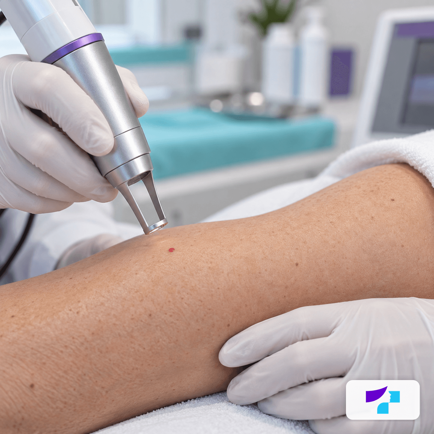
The visual shows laser removal of a small angioma-like vascular skin lesion in a dermatology clinic.
Laser treatment may be a good choice when the angioma is small, typical and the dermatologist is confident about the diagnosis. However, laser is not a universal solution for all red skin lesions. If histological examination is needed, surgical removal may be more appropriate.
After laser treatment there may be redness, slight swelling, darkening of the area or a small scab. In darker skin phototypes, the risk of pigmentation changes must be considered. In summer, laser procedures should be planned carefully because UV radiation may increase the risk of pigmentation after treatment.
Electrocoagulation
Electrocoagulation uses controlled heat. It may be suitable for small, raised angioma-like lesions.
A small scab usually forms after the procedure. It should not be picked or scratched, as this increases the risk of infection, pigmentation changes and scarring.
Cryotherapy
Cryotherapy means freezing the lesion with liquid nitrogen. It may be used in selected cases, but it is not always the first-choice method for angioma treatment.
After cryotherapy there may be redness, a blister, scab, lighter or darker pigmentation.
Shave excision or surgical removal
In some cases, an angioma can be shaved at skin level or surgically removed. This method is especially important if the lesion is atypical or histological examination is needed. More about similar procedures can be found in the section on skin lesion removal.
Surgical removal may leave a small scar. The risk of scarring depends on the size and location of the lesion, individual healing characteristics and aftercare.
What should you do if an angioma bleeds?
If an angioma starts bleeding after scratching or trauma, apply clean gauze or a dressing and press gently. Do not rub or scratch the lesion. If the bleeding does not stop, recurs or starts without clear trauma, see a doctor.
A bleeding lesion should not be burned, cut, treated with acids or handled with aggressive products at home.
Can angiomas be prevented?
There are no proven methods that reliably prevent cherry angiomas from forming. Their appearance is often related to age, genetics and individual skin characteristics.
However, overall skin health may benefit from:
- sun protection;
- avoiding skin trauma;
- regular skin self-examination;
- seeing a dermatologist if new or changing skin lesions appear.
How much does angioma removal cost?
The cost of angioma removal depends on:
- the type of angioma;
- its size;
- the number of lesions;
- the location;
- the chosen method;
- whether dermoscopy is needed;
- whether histological examination is needed;
- whether one or several lesions are removed.
Exact costs should be clarified during a consultation or according to the clinic’s price list.
Is angioma removal painful?
Discomfort depends on the method, the size of the angioma and its location. Small procedures are usually brief. Local anaesthesia may be used if needed.
After the procedure there may be tenderness, redness, swelling or a scab. Healing is usually short, but depends on the method and the individual reaction of the skin.
Possible risks and side effects
After angioma removal, possible effects include:
- redness;
- swelling;
- scab formation;
- temporary bleeding;
- infection;
- a lighter or darker spot;
- a small scar;
- incomplete removal;
- recurrence of the angioma;
- the need for histology if the diagnosis is unclear.
What should not be done with an angioma?
- Do not cut an angioma at home.
- Do not burn it with household methods.
- Do not use acids or caustic products without medical advice.
- Do not pick the scab after a procedure.
- Do not ignore rapid changes.
- Do not assume that every red skin lesion is a harmless angioma.
- Do not remove an atypical lesion without a diagnosis.
Angiomas in children and haemangiomas
Infantile haemangiomas are not the same as typical adult cherry angiomas. An infantile haemangioma is a benign vascular tumour that often appears in the first weeks of life. Many haemangiomas shrink over time, but not all should simply be observed without specialist assessment.
A medical examination is needed if the haemangioma:
- is located near the eyes, nose, lips or airways;
- is located in the genital area;
- ulcerates or bleeds;
- grows rapidly;
- is large, segmental or multiple;
- interferes with eating, vision, breathing or movement.
Vascular lesions in children should not be treated according to adult angioma principles without specialist evaluation.
Common myths about angiomas
Myth: every red dot on the skin is an angioma.
Not always. A red lesion may be another type of skin growth that requires different diagnosis.
Myth: an angioma is always dangerous.
A typical cherry angioma is usually not dangerous.
Myth: an angioma can turn into cancer.
A cherry angioma is not a precancerous lesion. However, it can be mistaken for other skin tumours.
Myth: angiomas always have to be removed.
If an angioma does not cause discomfort and the diagnosis is clear, treatment is often not necessary.
Myth: an angioma can be safely removed at home.
No. This can cause bleeding, infection, scarring and diagnostic error.
Myth: laser works for all red skin lesions.
Laser may be suitable for some angiomas, but the diagnosis must be known first.
Myth: there is never a scar after removal.
Any skin procedure can leave a mark, especially if the lesion is larger or healing is impaired.
Summary
Angiomas are most often benign vascular skin growths that appear as red dots or “red moles”. In adults, the most common type is a cherry angioma, which is usually harmless and does not always require treatment.
Angioma removal can be performed using laser treatment, electrocoagulation, cryotherapy or a surgical method. However, before removal it is important to confirm that the lesion really is an angioma. If it bleeds, changes, grows or looks atypical, the safest first step is a dermatologist’s examination and dermoscopy.
References and authoritative sources
- DermNet: patient and professional information about cherry angioma
- DermNet: information about venous lake as a benign vascular lesion
- DermNet: definition and development of infantile haemangioma
- MedlinePlus: patient information about cherry angioma
- Cleveland Clinic: symptoms, causes and treatment of cherry angioma
- Merck Manual Professional Edition: cherry angiomas as benign vascular skin lesions
- NCBI Bookshelf: medical overview of cherry haemangioma, also known as cherry angioma
