Trichoscopy
Trichoscopy is a non-invasive examination of the hair and scalp using a dermatoscope or a digital magnification system. It helps the doctor identify features of the hair shafts, follicular openings and scalp that may not be visible to the naked eye. The examination is used to diagnose different types of hair loss and scalp disorders, as well as to monitor treatment outcomes.
Trichoscopy is usually painless and does not require hair plucking or tissue sampling. However, it does not always provide a definitive diagnosis on its own. The findings are interpreted together with the patient’s symptoms, medical history and clinical examination and, if necessary, laboratory tests, tests for infection or a scalp biopsy.
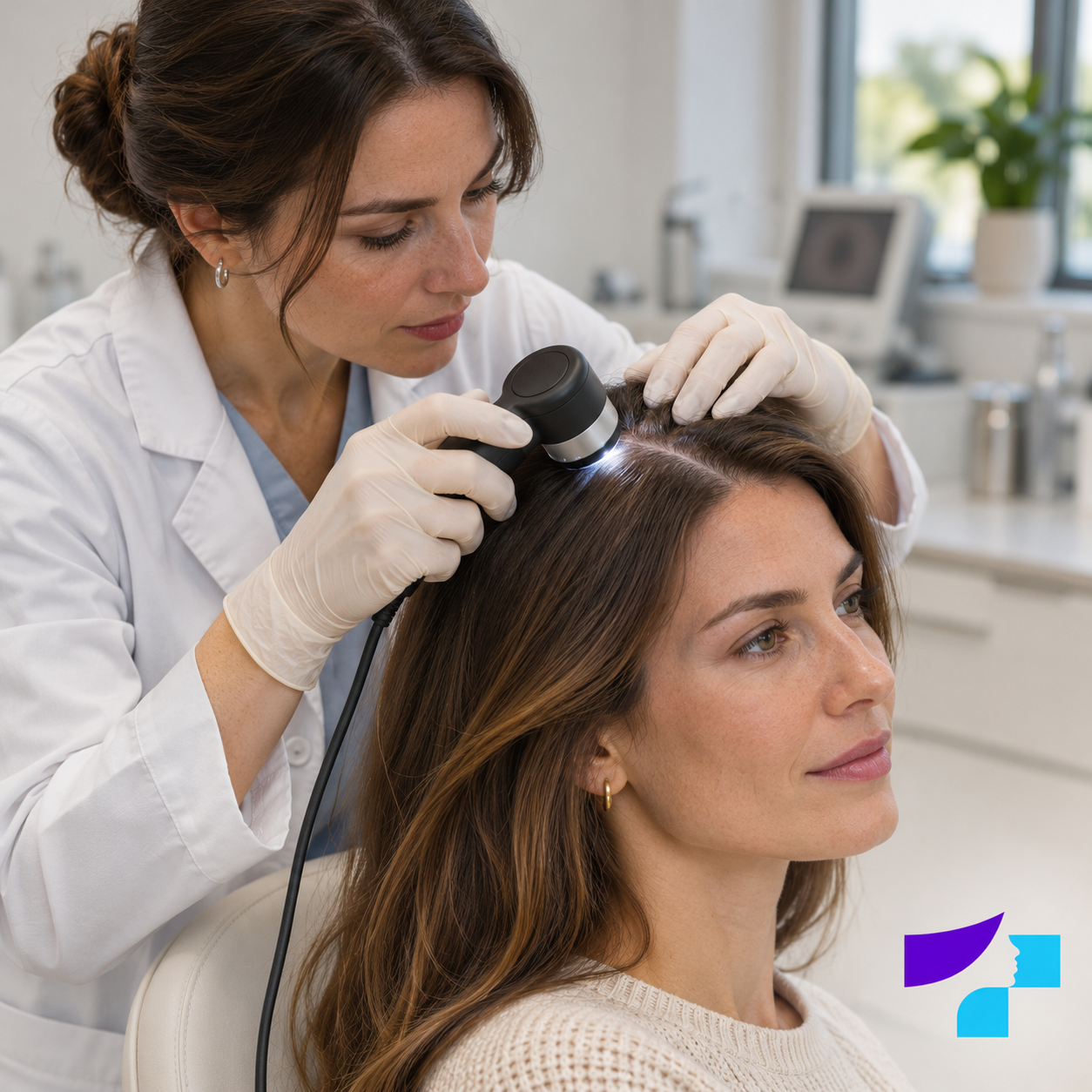
The image shows trichoscopy: the specialist parts the hair and examines the scalp under magnification with a dermatoscope. The examination is non-invasive and usually painless.
At a glance: key facts about trichoscopy
- Trichoscopy is a non-invasive diagnostic aid for examining the hair and scalp.
- The examination involves no incisions, injections or ionising radiation.
- Under magnification, the doctor assesses the hair shafts, follicular openings, skin around the follicles, scaling, colour changes and superficial blood vessels.
- The method can help distinguish between several types of alopecia and scalp disorders.
- A single trichoscopic feature does not always confirm one specific diagnosis.
- If supported by the system used, images can be saved and used to compare the condition at follow-up visits.
- Preparation requirements may vary, so follow the instructions provided by the clinic before your appointment.
Trichoscopy at the MedArt clinic
At MedArt, trichoscopy is used to assess hair loss and scalp problems. The examination may be performed during a consultation with a trichologist or as a separate diagnostic service, depending on the appointment and the patient’s needs.
Before your visit, it is advisable to confirm:
- whether you should book a separate trichoscopy appointment or a consultation with a trichologist;
- how to prepare your hair and scalp;
- how much time is allocated for the examination and explanation of the results;
- whether trichoscopy is included in the consultation fee or charged separately;
- the current price of the service.
MedArt’s currently published preparation guidance recommends not washing your hair or using styling products for 1–2 days before trichoscopy. If you receive different instructions when booking, follow those instructions, as preparation may depend on the purpose of the examination.
What is trichoscopy?
Trichoscopy is dermoscopy of the hair and scalp. A dermatoscope is an optical device that uses magnification and specialised lighting to reveal structures on the surface of the skin and hair.
The examination may be performed using:
- a handheld dermatoscope, through which the doctor directly examines selected areas;
- a videodermatoscope, which displays the image on a monitor;
- a digital trichoscopy system, which may allow images to be saved and compared and, in certain devices, measurements to be performed by software.
Digital trichoscopy does not always automatically include measurements of hair density, diameter or growth phases. This depends on the specific device, software and examination protocol. Therefore, “digital trichoscopy”, “computer-assisted trichoscopy” and “phototrichogram” are not fully interchangeable terms.
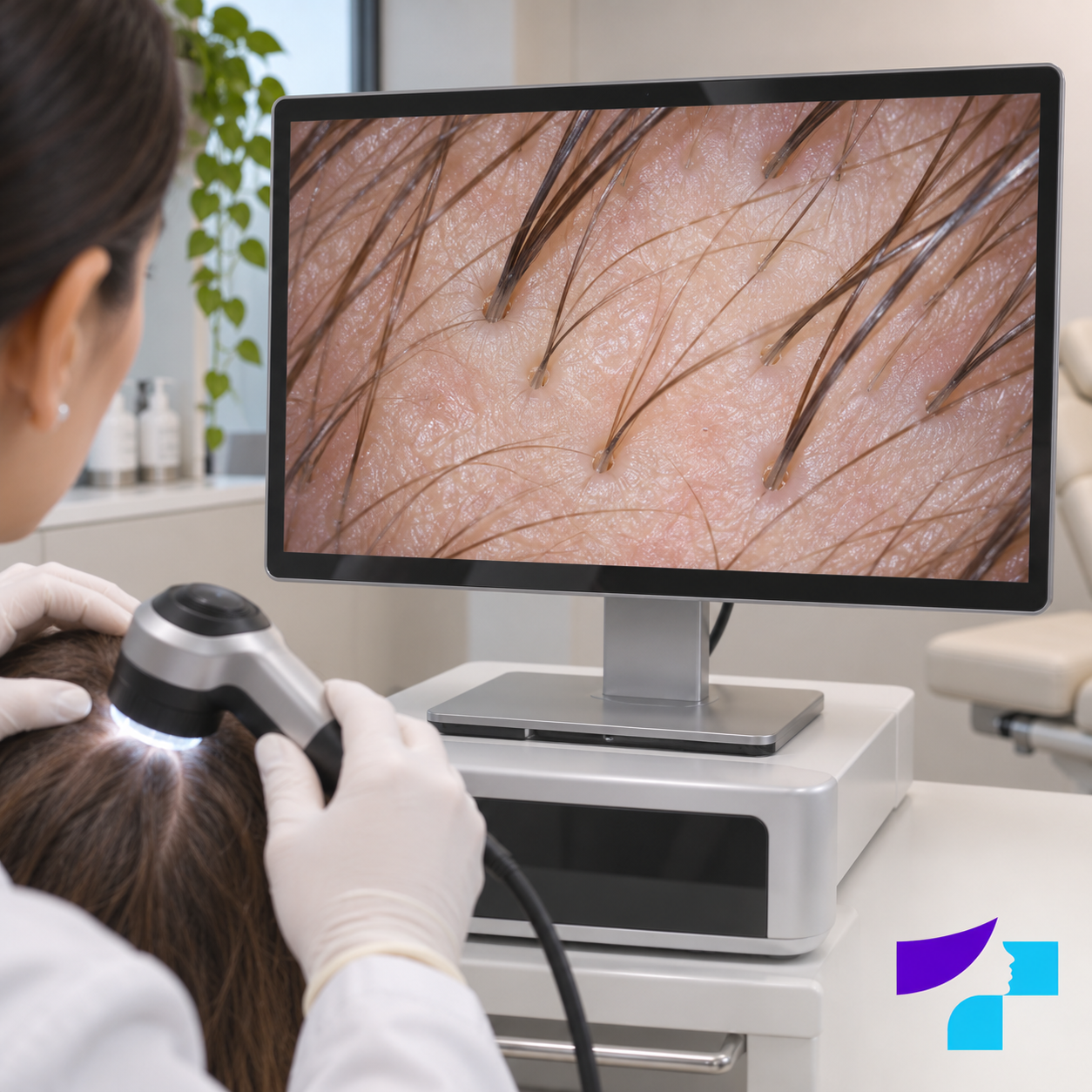
During digital trichoscopy, the magnified image is displayed on a monitor. The clinician assesses the hair shafts, follicular openings and superficial features of the scalp.
What can the doctor see during trichoscopy?
Trichoscopy generally assesses four main groups of structures:
- hair shafts;
- follicular openings;
- the skin around the hair follicles;
- the pattern of superficial blood vessels on the scalp.
Under magnification, the doctor may identify:
- variations in hair shaft diameter;
- very fine, short, broken or deformed hairs;
- changes in the number of hairs within follicular units;
- empty or enlarged follicular openings, openings filled with keratin or areas where openings are absent;
- redness or scaling around the follicles;
- diffuse scaling, crusts, pustules or discharge;
- changes in pigmentation or white and other coloured areas;
- distinctive patterns of superficial blood vessels;
- differences between several areas of the scalp.
Trichoscopy mainly shows the hair shafts, follicular openings and scalp surface. It does not allow direct examination of the entire hair follicle, or “hair root”, in the deeper layers of the skin. Standard trichoscopy is also not a direct measurement of scalp blood flow or the amount of sebum unless the particular device performs a separate, validated analysis.
When is trichoscopy recommended?
The examination may be useful when:
- more hair is shedding than usual;
- hair density or volume is gradually decreasing;
- the hair parting is widening;
- the hairline is receding;
- one or more hairless patches appear;
- hair breaks more easily or has become uneven in length;
- the scalp itches, hurts, burns, becomes red or scales;
- pustules, crusts or other signs of inflammation are visible;
- the doctor needs to distinguish between scarring and non-scarring hair loss;
- the most suitable site for a scalp biopsy needs to be selected;
- the baseline condition and response to treatment need to be documented.
Read more about the types of hair loss in the MedArt article Alopecia and hair loss.
Which conditions can trichoscopy help assess?
Trichoscopic findings are diagnostic clues, not an automatic diagnosis. The same feature may occur in several conditions, while the stage of the disease, previous treatment, hair characteristics and skin phototype can alter the appearance.
| Condition | What trichoscopy may help identify | Key limitation |
|---|---|---|
| Androgenetic alopecia | Variation in hair diameter, an increased proportion of fine hairs and changes in follicular units | The findings are interpreted together with the pattern and course of hair thinning and the clinical examination |
| Alopecia areata | Yellow and black dots, broken hairs, exclamation mark hairs and short regrowing hairs | Some features may also occur in other conditions |
| Telogen effluvium | Relatively uniform hair shaft diameter, empty follicular openings and short regrowing hairs of normal diameter. Findings are often non-specific. | The trichoscopic appearance may be non-specific; medical history is particularly important |
| Scarring alopecia | Loss of follicular openings, white areas, and redness or scaling around follicles | If suspected, a timely biopsy may be required |
| Hair shaft damage or mechanical hair pulling | Broken hairs of different lengths and characteristic hair shaft shapes | Hair-care habits and medical history must also be assessed |
| Fungal infection of the scalp | Broken, comma-shaped or corkscrew hairs and scaling | The infection often needs to be confirmed by microscopy, culture or another test |
| Psoriasis and seborrhoeic dermatitis | The distribution of scales and changes in skin colour and vascular patterns | Trichoscopy alone cannot always reliably distinguish between inflammatory conditions |
In a 2024 systematic review of androgenetic alopecia, the most frequently described trichoscopic features were variation in hair diameter, vellus or very fine hairs, and perifollicular changes. At the same time, the authors emphasised that no feature should be interpreted in isolation from the overall clinical picture.
When scarring alopecia is suspected, trichoscopy may help locate an active area suitable for biopsy. However, it does not replace tissue examination when histology is needed for an accurate diagnosis.
How is trichoscopy performed?
The exact course of the visit depends on the symptoms, the device used and whether trichoscopy is performed separately or as part of a consultation.
1. Discussion of symptoms
The specialist asks when the hair or scalp changes began, whether they developed suddenly or gradually, and how they have changed over time. Information about illnesses, surgery, fever, stress, weight changes, pregnancy, childbirth, diet, hair-care habits, medicines and supplements may be important.
2. Examination of the hair and scalp
The pattern of hair loss, hairline, density and condition of the scalp are assessed. If necessary, the doctor also examines the eyebrows, eyelashes, nails or other areas of the body.
3. Magnified examination
The dermatoscope is placed against the scalp or used in non-contact mode. A contact gel or another suitable liquid is sometimes used to improve image quality.
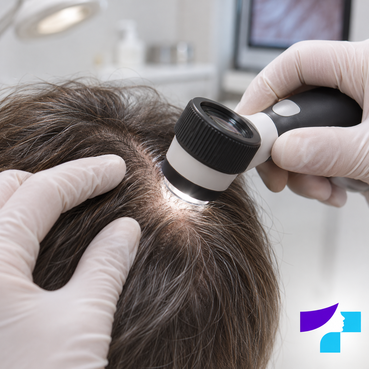
The specialist uses a dermatoscope to examine several areas of the scalp, noting hair shaft diameter, follicular openings, redness and scaling.
4. Comparison of several areas
The doctor may examine both the affected area and other areas for comparison. In cases of gradual hair thinning, it is often useful to compare the front and back of the scalp.
5. Saving images and explaining the findings
If supported by the system used, images can be saved for future comparison. The specialist explains the findings and discusses whether additional tests or treatment are needed.
The visible features can be assessed immediately, but the final conclusion may sometimes only be available after laboratory, mycological or biopsy results have been received.
How long does trichoscopy take?
Examining a single area may be brief, but the entire visit may take longer if a detailed discussion, photography of several areas, computer-assisted measurements or an explanation of the results is required.
The duration of the examination depends on:
- the number of areas being assessed;
- the method and device used;
- whether standardised images need to be saved;
- any additional tests;
- whether trichoscopy is part of a consultation.
Ask about the time allocated for your MedArt visit when making the appointment.
How should I prepare for trichoscopy?
There is no single preparation protocol for every trichoscopy method and clinic. Follow the instructions given by the particular healthcare provider before your visit.
MedArt currently recommends:
- not washing your hair for 1–2 days before the examination;
- not using hairspray, gel, mousse or other styling products.
Before your visit, it is also advisable to ask:
- whether dry shampoo, oils, tinted powders or hair fibres may be used;
- how long before the examination you should avoid dyeing your hair;
- what to do with medicated products applied to the scalp.
Do not stop prescribed medicines on your own solely because of the examination. If possible, bring previous laboratory and examination results, a list of medicines you use, and older photographs showing changes in your hair.
Is trichoscopy painful and safe?
Trichoscopy is non-invasive: it involves no incision, needle, tissue sampling or ionising radiation. Most people feel no pain during the examination, and no recovery time is required afterwards.
If the scalp is severely inflamed, painful or sensitive, contact with the device and parting the hair may cause brief discomfort. Tell the specialist about any pain or burning so that the examination can be adapted.
Trichoscopy can also be used to assess children’s hair and scalp. The need for the examination, the most appropriate specialist and its extent are determined according to the child’s age, symptoms and ability to cooperate.
Trichoscopy, trichogram or biopsy — what is the difference?
No single diagnostic method answers every question. Methods are selected according to the suspected diagnosis and may be combined.
| Method | How is it performed? | What does it help assess? | Main limitations |
|---|---|---|---|
| Trichoscopy | The hair and scalp are examined with a dermatoscope; no hairs are plucked | Hair shafts, follicular openings and superficial features of the scalp | Mainly superficial structures are visible; findings may be non-specific |
| Trichogram | A sample of hairs is plucked from a specific area and examined under a microscope | Features of hair roots and the hair growth cycle | May cause discomfort; sampling errors are possible |
| Phototrichogram | A standardised area, usually trimmed or shaved, is photographed repeatedly | Hair density, growth and, in some systems, growth phases | Repeated, precisely standardised images are required |
| Scalp biopsy | A small tissue sample is taken under local anaesthesia | The microscopic structure of the follicles and skin | Invasive; leaves a small wound and may result in a scar |
| Laboratory tests | A blood sample or another biological sample is collected | Deficiencies and endocrine, inflammatory, infectious or other systemic factors | Do not show the pattern of hair loss on the scalp |
What can trichoscopy not determine on its own?
On its own, trichoscopy:
- does not always identify the sole cause of hair loss;
- does not confirm iron or other nutrient deficiencies;
- does not confirm thyroid disease or hormonal disorders;
- does not always reliably rule out a fungal or bacterial infection;
- does not replace a biopsy when a scarring or unclear process is suspected;
- cannot guarantee the outcome of a particular treatment;
- is not a treatment procedure;
- is not a basis for independently starting prescription medicines or high doses of supplements.
A person may have several causes of hair loss at the same time. A reliable diagnosis therefore relies on all available information.
When are laboratory tests or a biopsy needed?
Additional examinations are selected according to the individual clinical question.
- Blood tests may be needed if the history or examination suggests anaemia, iron deficiency, thyroid dysfunction, nutrient deficiency, hormonal changes or another systemic cause.
- Mycological testing may be required for scaly patches, broken hairs or other signs of a possible fungal infection.
- Microbiological testing may be useful if pustules, discharge or signs of a bacterial infection are present.
- Scalp biopsy may be needed if scarring alopecia is suspected or the diagnosis remains unclear after clinical examination and trichoscopy.
Not every patient needs the same set of tests.
What does a trichoscopy result mean?
A trichoscopy result is usually not a single number or a simple “positive/negative” conclusion. The specialist builds an overall picture from several features and different areas of the scalp.
Several conclusions are possible after the examination:
- the findings, together with the clinical picture, are consistent with a particular diagnosis;
- the range of possible diagnoses has been narrowed, but laboratory or infection testing is needed;
- there are features that indicate a biopsy is required;
- there are no specific features, making the course of the condition and other examinations particularly important;
- more than one mechanism of hair loss may be present at the same time.
If the diagnosis is sufficiently clear, the treatment plan may be discussed during the same visit. If the information is insufficient, it is safer to wait for the results of additional examinations first.
When is trichoscopy repeated?
Repeat trichoscopy can help determine whether the condition is progressing, stabilising or improving. Comparison is more reliable when the same areas are photographed again with a similar parting position, hair length, magnification and lighting.
There is no single follow-up interval for every patient. It depends on:
- the diagnosis;
- disease activity;
- the treatment used;
- how soon objectively assessable changes can be expected.
The hair growth cycle is slow, so an assessment performed too early may not show whether treatment is effective.
When should you see a specialist?
A routine consultation is recommended if hair shedding remains greater than usual for a prolonged period, hair density gradually decreases, the parting widens, the hairline recedes, or scalp itching and scaling repeatedly occur.
More prompt medical assessment is needed if:
- hair loss begins suddenly or progresses very rapidly;
- round or irregular hairless patches appear;
- there is pain, burning or marked scalp sensitivity;
- redness, thick scaling, crusting or bleeding is visible;
- there are pustules, pus-filled lesions or discharge;
- areas of skin become smooth or shiny, raising suspicion of scarring;
- the eyebrows, eyelashes or body hair are also lost;
- hair loss is accompanied by fever, pronounced weakness, unexplained weight changes or other general symptoms.
Early diagnosis is particularly important when scarring alopecia is possible, because hair may not regrow where follicles have been destroyed.
Myths and facts about trichoscopy
Myth: trichoscopy treats hair loss
Fact: it is a diagnostic and monitoring method. Treatment is selected according to the diagnosis.
Myth: one magnified image always provides an exact diagnosis
Fact: the combined interpretation of several areas and features, medical history and sometimes additional examinations are important.
Myth: trichoscopy and a trichogram are the same
Fact: hairs are not plucked during trichoscopy; a trichogram requires a sample of plucked hairs for microscopic examination.
Myth: everyone with hair loss needs the same tests
Fact: tests are selected according to the symptoms, medical history and suspected diagnosis.
Myth: trichoscopy guarantees that lost hair can be fully restored
Fact: the prognosis depends on the diagnosis, duration of the process, condition of the follicles and available treatment options.
Myth: all hair loss can be explained by a vitamin deficiency
Fact: hair loss has many possible causes, and supplements help only in specific situations.
Summary
Trichoscopy is a non-invasive diagnostic aid for examining the hair and scalp. It helps identify and document features of the hair shafts, follicular openings and scalp, distinguish between several types of hair loss, select a biopsy site and monitor changes over time.
The most reliable result is achieved when the trichoscopic appearance is assessed together with the patient’s symptoms, medical history, clinical examination and, where necessary, targeted additional investigations.
Medical disclaimer
This information is for educational purposes and does not replace an individual consultation, diagnosis or treatment by a trichologist or dermatologist. Trichoscopic findings must be assessed together with symptoms, medical history and other investigations. Seek medical advice if hair loss is sudden, patchy, rapidly progressive or accompanied by pain, redness, pronounced scaling, pustules or possible signs of scarring.
References and authoritative sources
- DermNet: overview of trichoscopy
- European expert consensus statement on current issues in trichology
- Journal of Clinical Medicine: systematic review of trichoscopy in the diagnosis of androgenetic alopecia
- International Dermoscopy Society task force: systematic review of hair and scalp trichoscopy in people with skin of colour
- American Academy of Dermatology: diagnosis and treatment of hair loss
- DermNet: trichoscopy of localised cicatricial alopecia
- American Academy of Dermatology: diagnosis and treatment of frontal fibrosing alopecia
- International Journal of Trichology: advantages and limitations of hair evaluation methods
Content author
Frequently Asked Questions
1. Is trichoscopy painful?
Usually not. The device touches the scalp or is used in non-contact mode. If the skin is inflamed or very sensitive, light contact may cause brief discomfort.
2. Can you wash your hair before trichoscopy?
Follow the instructions of the specific clinic. MedArt currently recommends not washing your hair for 1–2 days before the examination.
3. Can you use dry shampoo or hairspray before the appointment?
It is usually recommended not to use them because product residue may obscure the scalp and hair shafts or resemble diagnostic signs.
4. Can you dye your hair before trichoscopy?
Dye residue may interfere with the examination. It is advisable to ask about a suitable interval after hair colouring when booking the appointment.
5. Does trichoscopy identify the cause of hair loss?
It can provide important diagnostic clues, but medical history, clinical examination, laboratory tests, infection testing or a biopsy may also be needed.
6. Are trichoscopy and a trichogram different?
Yes. Trichoscopy is a magnified examination without pulling out hairs. For a trichogram, a specific sample of hairs is plucked and the hair roots and signs of the growth cycle are evaluated under a microscope.
7. Can trichoscopy be performed on children?
Yes, the method is non-invasive and may be suitable for children. A specialist assesses the need for the examination and its scope.
8. Does hair need to be shaved for trichoscopy?
Usually not for standard trichoscopy. Trimming or shaving a small area may be required for a phototrichogram or another standardised hair growth analysis.
9. Is the result available immediately?
The specialist can assess the visible findings during the appointment. If laboratory tests, a mycological examination or a biopsy are needed, the final conclusion will be available later.
10. Is a referral required for trichoscopy?
It depends on the healthcare institution and the payment arrangements for the service. MedArt is a private healthcare centre, so it is advisable to confirm the booking conditions before the appointment.
11. How much does trichoscopy cost?
The price depends on the clinic, the method used and whether the examination is a separate service or part of a consultation. Check the current MedArt price in the price list or confirm it when booking.
12. Is treatment determined immediately after trichoscopy?
If the clinical picture and findings are sufficient, the treatment plan can be discussed during the same appointment. In an unclear case, additional examinations are needed first.
Consultation fee
| Qualification | Clients of the clinic | First visit |
|---|---|---|
| Trichoscopy | 57 € | 60 € |
Trichoscopy prices
| Service | Price |
|---|---|
| Trichoscopy | EUR 27 |





 +1
 +1
 +1 242
 +1 246
 +1 268
 +1 441
 +20
 +212
 +213
 +216
 +218
 +220
 +221
 +222
 +223
 +223
 +224
 +225
 +226
 +227
 +228
 +229
 +230
 +231
 +232
 +234
 +235
 +236
 +237
 +238
 +239
 +239
 +240
 +241
 +242
 +243
 +244
 +245
 +248
 +249
 +250
 +251
 +252
 +253
 +254
 +255
 +256
 +257
 +258
 +260
 +261
 +262
 +263
 +264
 +265
 +266
 +267
 +268
 +269
 +269
 +27
 +284
 +290
 +291
 +297
 +298
 +299
 +30
 +31
 +32
 +33
 +34
 +340
 +345
 +350
 +351
 +352
 +353
 +354
 +355
 +356
 +358
 +359
 +36
 +370
 +371
 +372
 +373
 +374
 +375
 +376
 +377
 +378
 +380
 +385
 +386
 +387
 +389
 +39
 +39
 +40
 +41
 +41
 +420
 +421
 +43
 +45
 +46
 +47
 +473
 +48
 +49
 +500
 +501
 +502
 +503
 +504
 +505
 +506
 +507
 +509
 +51
 +52
 +53
 +54
 +55
 +56
 +57
 +58
 +590
 +591
 +593
 +594
 +594
 +595
 +596
 +597
 +598
 +60
 +61
 +62
 +63
 +64
 +649
 +65
 +66
 +664
 +671
 +672
 +672
 +672
 +673
 +674
 +675
 +676
 +677
 +678
 +679
 +680
 +681
 +682
 +683
 +684
 +686
 +687
 +688
 +689
 +690
 +691
 +692
 +6982
 +7
 +7
 +7
 +7
 +758
 +767
 +784
 +809
 +809
 +81
 +82
 +84
 +850
 +852
 +853
 +856
 +86
 +868
 +869
 +876
 +880
 +885
 +886
 +90
 +91
 +92
 +93
 +94
 +95
 +960
 +961
 +962
 +963
 +964
 +965
 +967
 +968
 +971
 +972
 +973
 +974
 +975
 +976
 +977
 +98
 +993
 +994
 +995
 +996
 +996