Mole removal may be needed for medical, functional or aesthetic reasons. A mole may rub against clothing, be repeatedly injured during shaving, cause discomfort or bother the patient visually. In some cases, the doctor may recommend excising the lesion so that the tissue can be sent for histological examination and the diagnosis can be clarified.
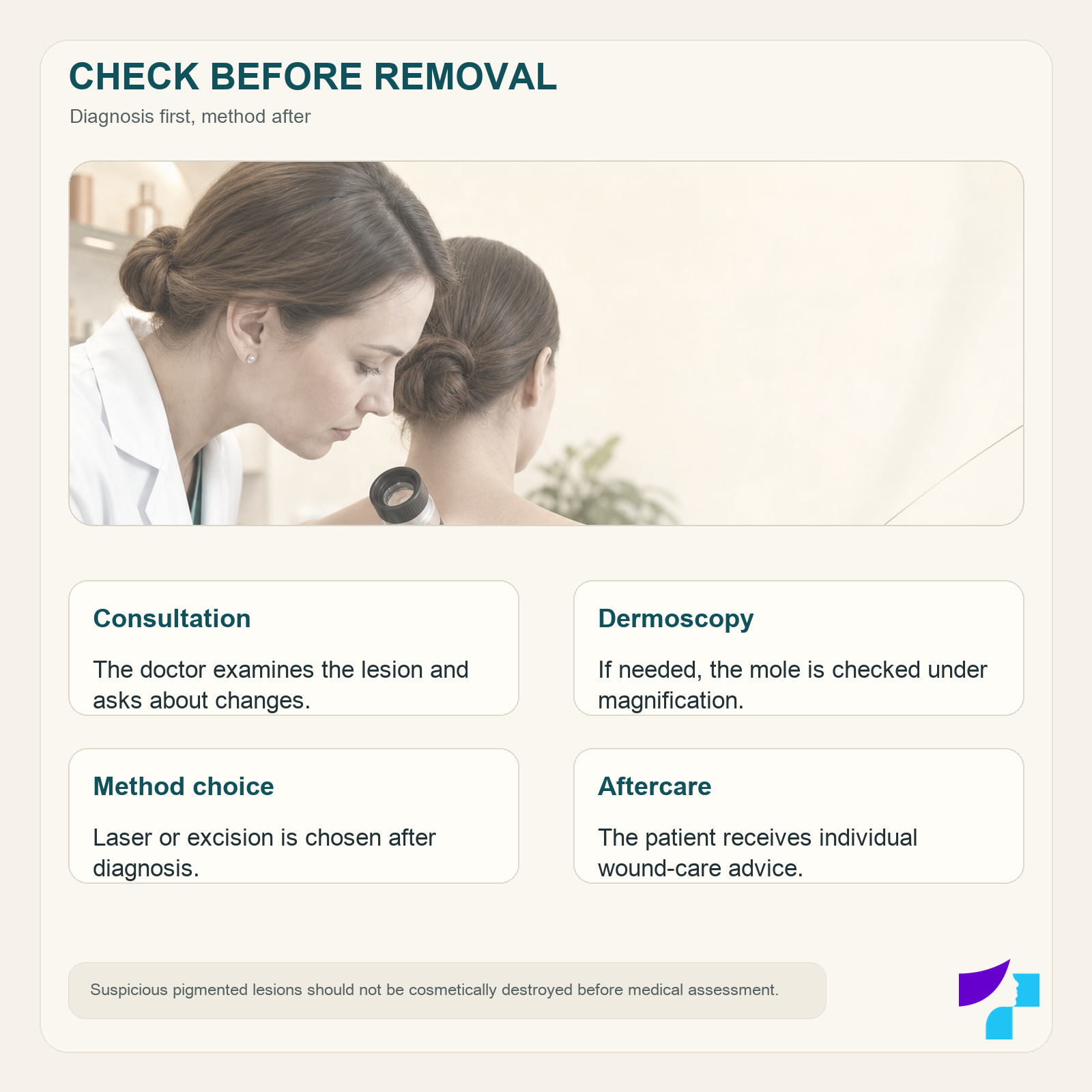
Before the procedure, the mole should be examined by a dermatologist. The doctor assesses its appearance, structure and changes over time and, if necessary, performs a mole examination with a dermatoscope. Only after diagnostic assessment can it be decided whether the lesion should be monitored, removed surgically or, in selected cases, treated with laser.
In everyday language, patients may call many different pigmented and non-pigmented skin lesions “moles”. A melanocytic naevus, seborrhoeic keratosis, papilloma, wart and angioma are not the same diagnosis, and their treatment may differ. The precise diagnosis and the most appropriate removal method are determined by the doctor.
Important in brief
- A mole should be checked by a dermatologist before removal.
- Not all moles are suitable for laser removal.
- If the lesion is suspicious, the method should allow tissue to be obtained for histological examination.
- Surgical excision and many laser procedures are performed under local anaesthesia.
- A scar or pigmentation changes may remain after any removal method.
- The exact method, extent and cost of the procedure can be determined only after the lesion has been examined.
When should a mole be removed?
Most moles are benign and do not need to be removed. A dermatologist may recommend mole removal if the lesion:
- regularly rubs against clothing, jewellery or equipment;
- is repeatedly injured during shaving, combing or sports;
- bleeds, hurts, itches or becomes inflamed repeatedly;
- causes functional discomfort;
- causes significant aesthetic concern;
- looks suspicious and histology is needed to clarify the diagnosis;
- in the doctor’s opinion, should not be left for observation only.
Repeated trauma does not automatically mean that a mole must be excised. The doctor assesses the diagnosis, location, frequency of injury and the expected result after the procedure.
If a mole changes in size, shape or colour, becomes asymmetric, starts bleeding or ulcerating, or looks clearly different from other lesions, diagnostic assessment is needed first. Such changes do not always mean a malignant process, but they should not be ignored. Read more about signs that deserve attention in the article “Birthmarks and moles”.
Mole examination before removal
Mole examination before removal is important because the procedure method is determined not only by the lesion’s size or the patient’s preference, but also by the possible diagnosis.
During the visit, the dermatologist may:
- ask how long the lesion has been present and whether it has changed;
- ask about bleeding, itching, pain and trauma;
- examine the specific mole and the surrounding skin;
- assess other skin lesions;
- perform dermoscopy;
- compare the lesion with previous images, if available;
- recommend observation, biopsy or complete excision.
Dermoscopy is a non-invasive examination method that allows the doctor to assess pigment distribution, blood vessels and other structures under magnification, including signs that are not sufficiently visible to the naked eye.
Dermoscopy helps the doctor decide on further management, but in suspicious lesions the final diagnosis is provided by histological examination of tissue. If a mole has suspicious features, it is usually removed in a way that allows the obtained material to be examined in a laboratory.
A suspicious pigmented lesion should not be cosmetically destroyed with laser, cryotherapy or another destructive method before the doctor has assessed whether histology is needed. If the tissue is destroyed during the procedure, a complete laboratory examination afterwards is not possible.
Can a mole be removed during the same visit?
Sometimes a checked lesion that appears benign according to the doctor’s assessment can be removed during the same visit. However, this is not guaranteed.
The procedure may need to be planned separately if:
- a more extensive surgical excision is needed;
- histological examination must be planned;
- the lesion is located in a complex anatomical area;
- several moles need to be removed;
- the patient has health conditions or medications that must be considered;
- the procedure requires more time or special preparation.
The possibility of examination and removal during one appointment should be discussed with the clinic when booking the visit.
Mole removal methods
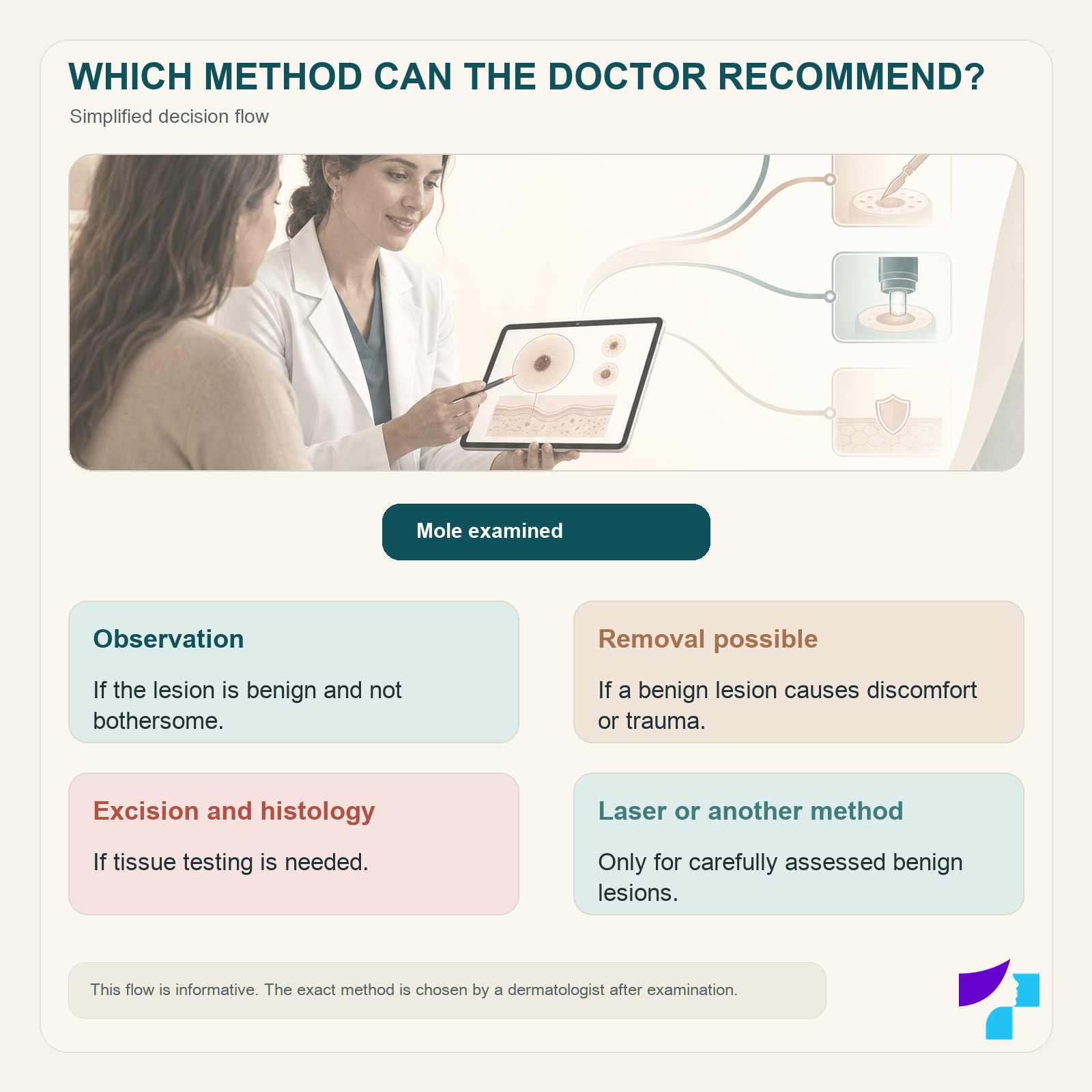
The dermatologist chooses the removal method by considering:
- the lesion’s diagnosis;
- its size and depth;
- location;
- pigmentation pattern;
- suspicious features;
- the need for histology;
- the expected risk of scarring;
- the patient’s health status;
- previous treatment or trauma to the lesion.
The main methods are surgical excision, superficial surgical removal and, in selected carefully assessed cases, laser treatment.
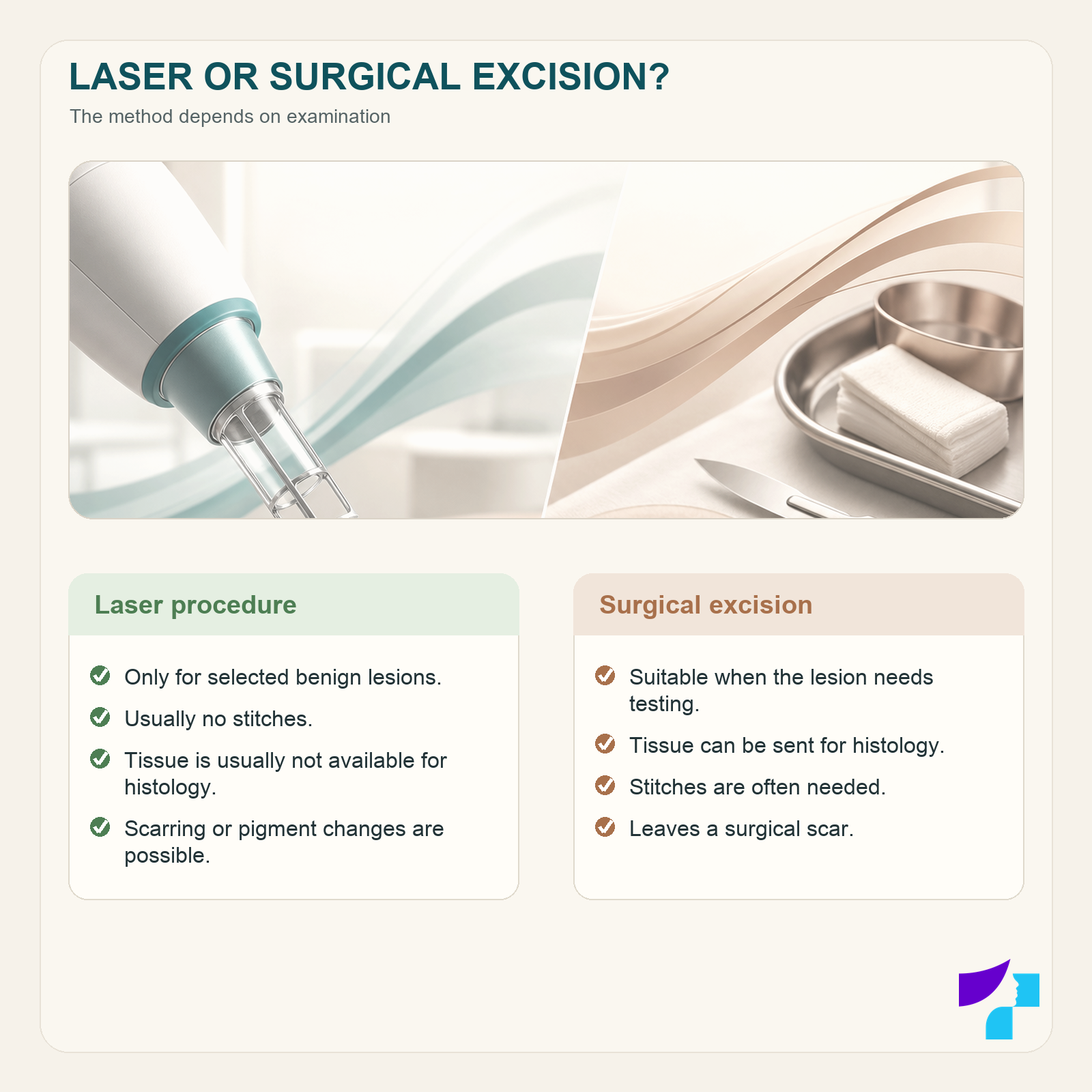
| Criterion | Laser procedure | Surgical excision |
|---|---|---|
| Suitability | For selected benign lesions approved by the doctor | For benign and suspicious lesions according to the doctor’s decision |
| Tissue for histology | Destroyed tissue is usually not available for a complete examination | The removed lesion can be sent for histology |
| Local anaesthesia | May be needed | Usually needed |
| Stitches | Usually not needed | Often needed |
| Healing pattern | Superficial wound or crust formation | Surgical wound healing with or without stitches |
| Scar risk | A scar or pigmentation changes are possible | A surgical scar remains |
| Chance of recurrence | Possible if lesion cells remain deeper in the skin | Lower after complete excision, but depends on the diagnosis and procedure |
Laser mole removal
Laser mole removal may seem simpler and less invasive than surgical excision, but laser is not a universal method and is not suitable for every pigmented lesion.
Laser treatment can be considered only after the doctor has assessed the lesion and concluded that destroying it will not interfere with the necessary diagnostics. Melanocytic naevi and lesions with suspicious features require particular caution.
Patients also often ask about mole removal with a CO₂ laser. However, the device name alone does not determine whether the procedure is suitable. The decision depends on the diagnosis, depth, location and need for histology.
Before laser treatment, the skin may be numbed with local anaesthesia. After the procedure, a superficial wound or crust may form in the treated area. During healing, the following may occur:
- redness;
- tenderness;
- a lighter or darker patch;
- a slight depression or thickening of the skin;
- a scar;
- recurrent pigmentation.
Laser treatment cannot guarantee a scar-free result. In addition, once the tissue has been destroyed, it is usually not possible to obtain complete material for histological examination.
Surgical mole excision
Surgical mole removal, or excision, is a procedure during which the doctor cuts out the skin lesion. Depending on the diagnosis, only the lesion itself or a small rim of surrounding tissue may be removed.
The procedure is usually performed under local anaesthesia. The patient remains awake, but sharp pain should not be felt in the numbed area during the procedure. After the lesion is removed, the wound may be closed with one or more layers of stitches. Some small or superficial wounds may be left to heal without stitches.
An important advantage of surgical excision is that the removed tissue can be preserved and sent for histological examination. In the laboratory, a pathologist examines the lesion’s cells under a microscope, clarifies the diagnosis and, if necessary, assesses whether the lesion has been removed completely.
Surgical excision leaves a scar. The doctor plans the procedure so that the scar is as discreet as possible, but the result is also affected by the lesion’s size, location, skin tension and the patient’s individual healing response.
Superficial or shave excision
In selected cases, a raised lesion that appears benign according to the doctor’s assessment can be removed with tangential, or shave, excision. During the procedure, the part of the lesion that protrudes above the skin is removed with a surgical instrument.
This method does not always remove all deeper cells, so the lesion or pigmentation may appear again. Depending on the clinical situation, the obtained tissue can be sent for histological examination.
Other skin lesion removal methods
Electrosurgery, cryotherapy and other methods may be used to treat certain benign skin lesions, but they are not universal methods for removing melanocytic moles.
A lesion that the patient considers to be a mole may turn out to be:
- seborrhoeic keratosis;
- papilloma;
- wart;
- angioma;
- sebaceous gland hyperplasia;
- another benign or malignant skin lesion.
For this reason, the diagnosis should be determined first, and only then should the procedure be chosen.
How to prepare for mole removal?
Before the procedure, the doctor should be informed about:
- regularly used medicines and supplements;
- medicines that affect blood clotting;
- allergies, including reactions to anaesthetic agents;
- blood clotting disorders;
- diabetes and other chronic diseases;
- a pacemaker or other implanted devices;
- pregnancy or breastfeeding;
- previous excessive or keloid scarring.
Medicines prescribed by a doctor, including blood-thinning medication, should not be stopped independently. Any necessary changes must be decided by the treating doctor.
Before the visit, the mole should not be cut, burned, tied off or treated with acids or other home remedies. On the day of the procedure, it is advisable not to apply cosmetics to the treated area unless the doctor has instructed otherwise.
How does mole removal work?
The exact course of the procedure depends on the chosen method, but it usually includes several steps.
1. Dermatologist consultation
The doctor asks how long the lesion has been present, whether it has changed, been injured, bled or caused other symptoms.
2. Lesion assessment
The dermatologist examines the mole and, if necessary, performs dermoscopy. If the lesion is suspicious, a method is chosen that allows tissue to be obtained for histological examination.
3. Method selection
The doctor determines whether surgical excision, superficial removal, laser treatment or another treatment is more appropriate. The patient is informed about the expected result, scar risk, healing and aftercare.
4. Local anaesthesia
Before surgical removal and some laser procedures, the treatment area is numbed. A brief sting or burning sensation may be felt during the injection.
5. Lesion removal
The doctor removes the mole using the method chosen in advance. The duration of the procedure depends on the number, size and location of the lesions and the complexity of the procedure.
6. Wound closure and dressing
After surgical excision, stitches, wound closure strips and a dressing may be applied. After laser treatment or superficial removal, the wound may be left to heal without stitches.
7. Histological examination
If needed, the removed tissue is sent to the laboratory. The clinic explains how and when the histology results will be provided.
8. Instructions after the procedure
The patient receives individual instructions on dressing changes, washing, physical activity, stitch removal and what to do if complications occur.
Does mole removal hurt?
Mole removal is usually performed with local anaesthesia. Most often, a brief sting and burning sensation are felt when the anaesthetic is injected. Once the area is numb, the patient may feel touch or pressure during the procedure, but should not feel pain.
After the anaesthetic wears off, tenderness, a pulling sensation or mild pain may occur. The intensity depends on the extent of the procedure and the body area.
The doctor should be contacted if pain does not gradually decrease but becomes stronger, or if increasing redness, heat, swelling or discharge from the wound appears.
Histological examination after mole removal
Histological examination is the microscopic examination of removed tissue. It is performed by a pathologist to determine what cells make up the tissue sample and whether benign, atypical or malignant changes are present.
Histology may be needed to:
- confirm the precise diagnosis;
- rule out or confirm a malignant skin tumour;
- assess whether the lesion has been removed completely;
- determine whether additional excision is needed;
- plan further monitoring or treatment.
A doctor’s examination and dermoscopy help assess the risk of a lesion, but the final diagnosis of a suspicious melanocytic lesion requires histological examination.
If the lesion is destroyed with laser, cryotherapy or another destructive method, a complete tissue sample may not be available after the procedure. Therefore, the decision about the removal method must be made before the procedure.
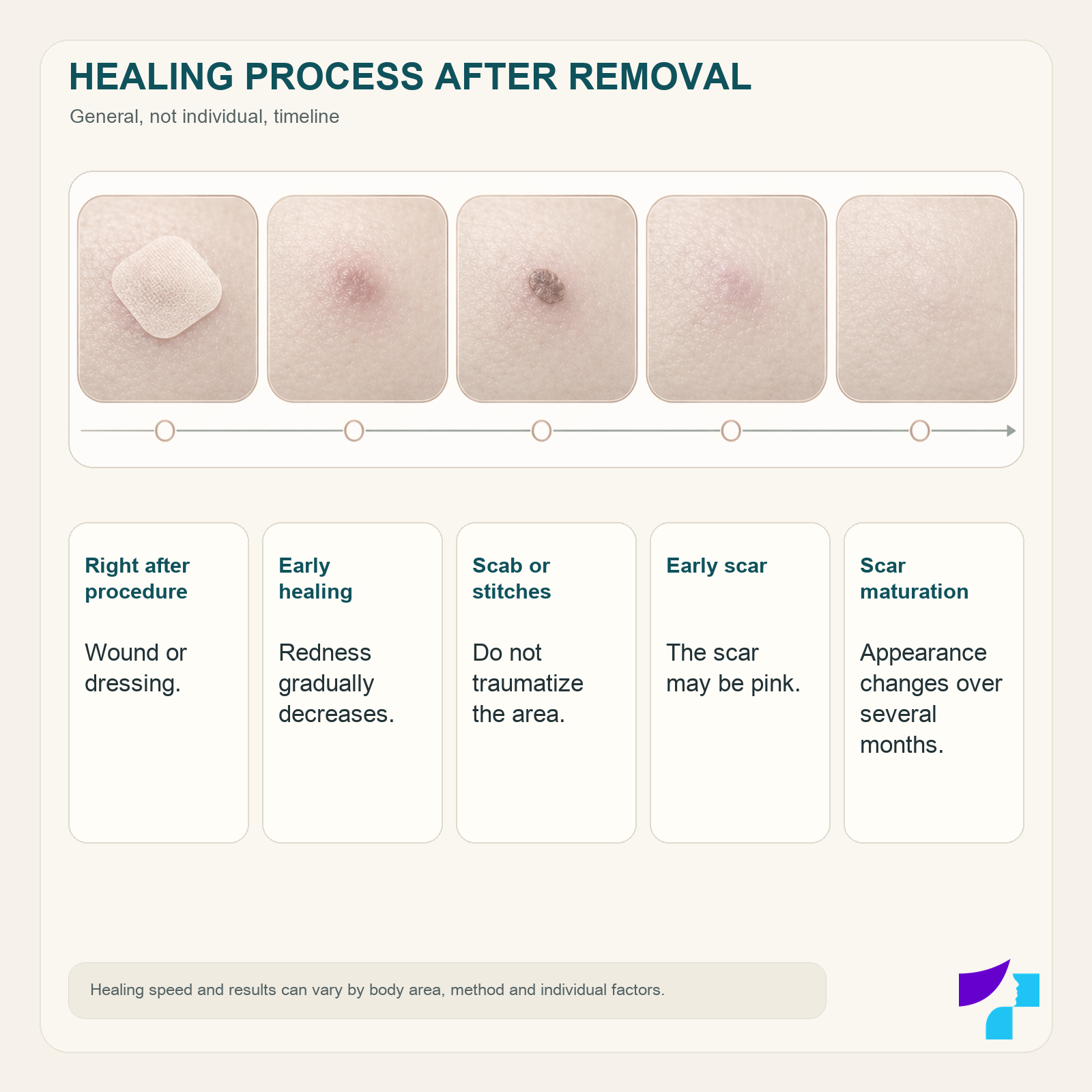
Healing and skin care after mole removal
Wound care after mole removal depends on the method used, wound size, stitches and body area. The instructions given by the doctor who performed the procedure should always be followed.
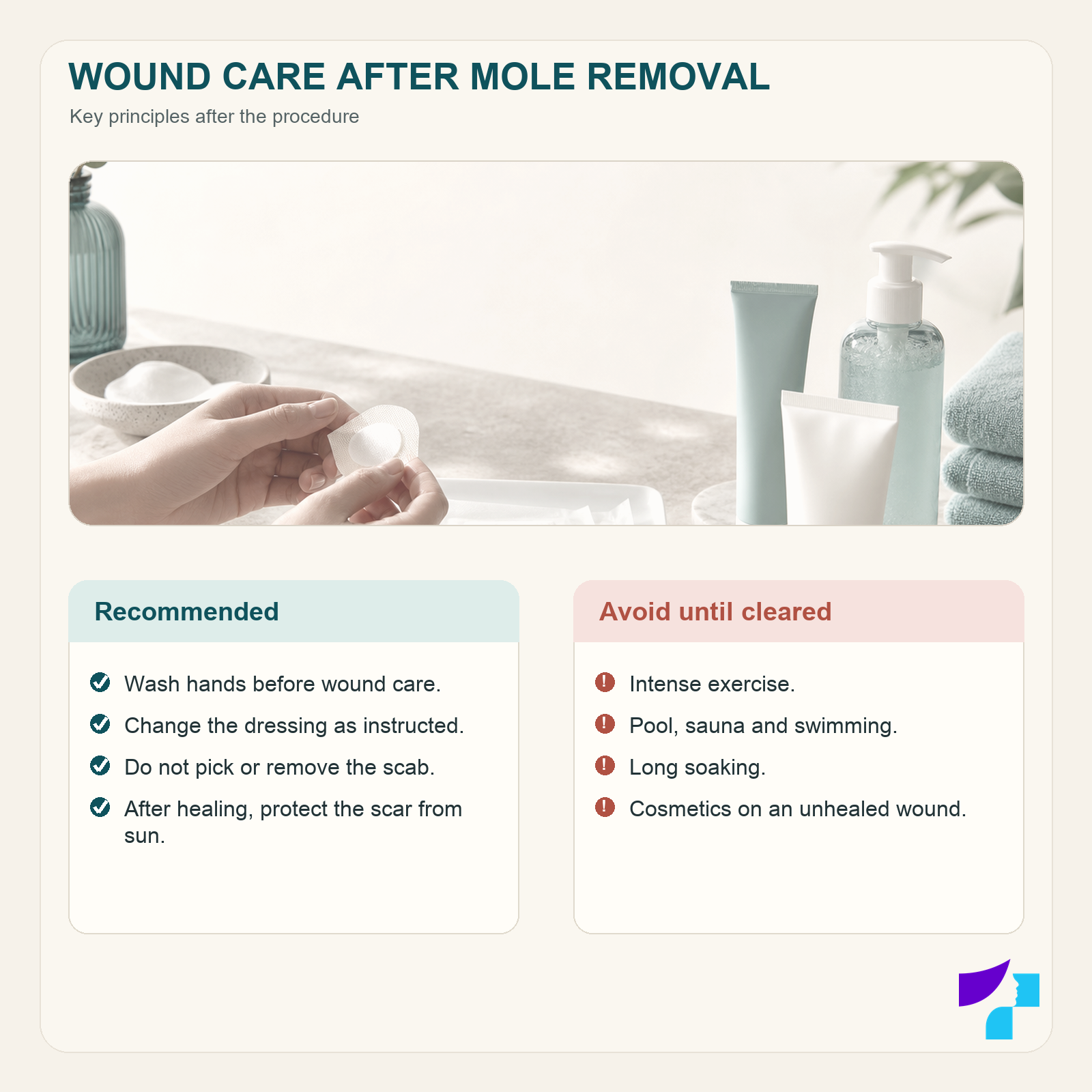
General recommendations may include:
- washing hands before touching the wound;
- keeping the initial dressing for the time specified by the doctor;
- cleaning the wound only with the recommended products;
- gently drying the area after washing instead of rubbing it;
- using a doctor-recommended ointment and a clean dressing if needed;
- not removing the crust;
- not applying cosmetics to an unhealed wound;
- not using antibiotic ointments without the doctor’s instruction;
- avoiding clothing and movements that rub or stretch the wound;
- attending follow-up or stitch removal at the specified time.
Different procedure wounds are cared for differently. In some cases, the doctor may recommend keeping the wound clean, moist with a suitable product and covered, while in other situations the instructions may differ.
Washing after the procedure
The doctor determines when the wound may be made wet. Once washing is allowed, the area is usually washed gently and dried carefully without rubbing.
Until the wound has closed, it should not be soaked for a long time in a bath, swimming pool or other body of water.
Exercise after mole removal
Intense physical activity may need to be limited after the procedure because sweating, skin tension and increased pressure in the wound area may promote bleeding or wound edge separation.
The duration of restrictions depends on the extent and location of the procedure. A wound on the back, shoulder, joint or another mobile area may require longer protection.
Sauna and swimming pool
Sauna, swimming pool and swimming should be postponed until the wound has closed and the doctor has allowed these activities. Moisture, heat, sweating and microorganisms may increase the risk of irritation or infection.
Sun exposure and SPF
A fresh wound and a healing scar should not be exposed to direct sun. After the skin has healed, the scar should be protected with clothing and a broad-spectrum sunscreen. Sun exposure may contribute to persistent pigmentation changes and make the scar more visible.
Will there be a scar after mole removal?
Any procedure that affects deeper layers of the skin may leave a scar. It is not possible to excise a skin lesion completely without any scar, but careful technique and proper wound care can help make the scar less noticeable.
The appearance of the scar is influenced by:
- the removal method used;
- the size and depth of the lesion;
- the body area;
- skin tension;
- the patient’s age and skin characteristics;
- smoking;
- infection;
- repeated trauma to the wound;
- sun exposure;
- a tendency to hypertrophic or keloid scars.
After laser treatment, a classic linear surgical scar may not be present, but a round lighter or darker area, depression, thickening or another change in skin texture may occur.
At first, the scar may be pink, firmer or slightly raised. Its colour and texture may continue to change for several months. Silicone products, massage or other scar care products should be discussed with the doctor after the wound has fully closed.
Can a mole grow back after removal?
A mole or pigment may appear again at the removal site if lesion cells remain in the deeper layers of the skin. This is more likely after superficial removal or a procedure whose aim was not complete excision.
Recurrent pigment does not always mean a malignant process, but it should not be ignored. Changes in a scar can be more difficult to interpret than the original lesion, so a dermatologist’s examination is needed.
The removal site should not be burned, cut or treated again at home.
Possible side effects
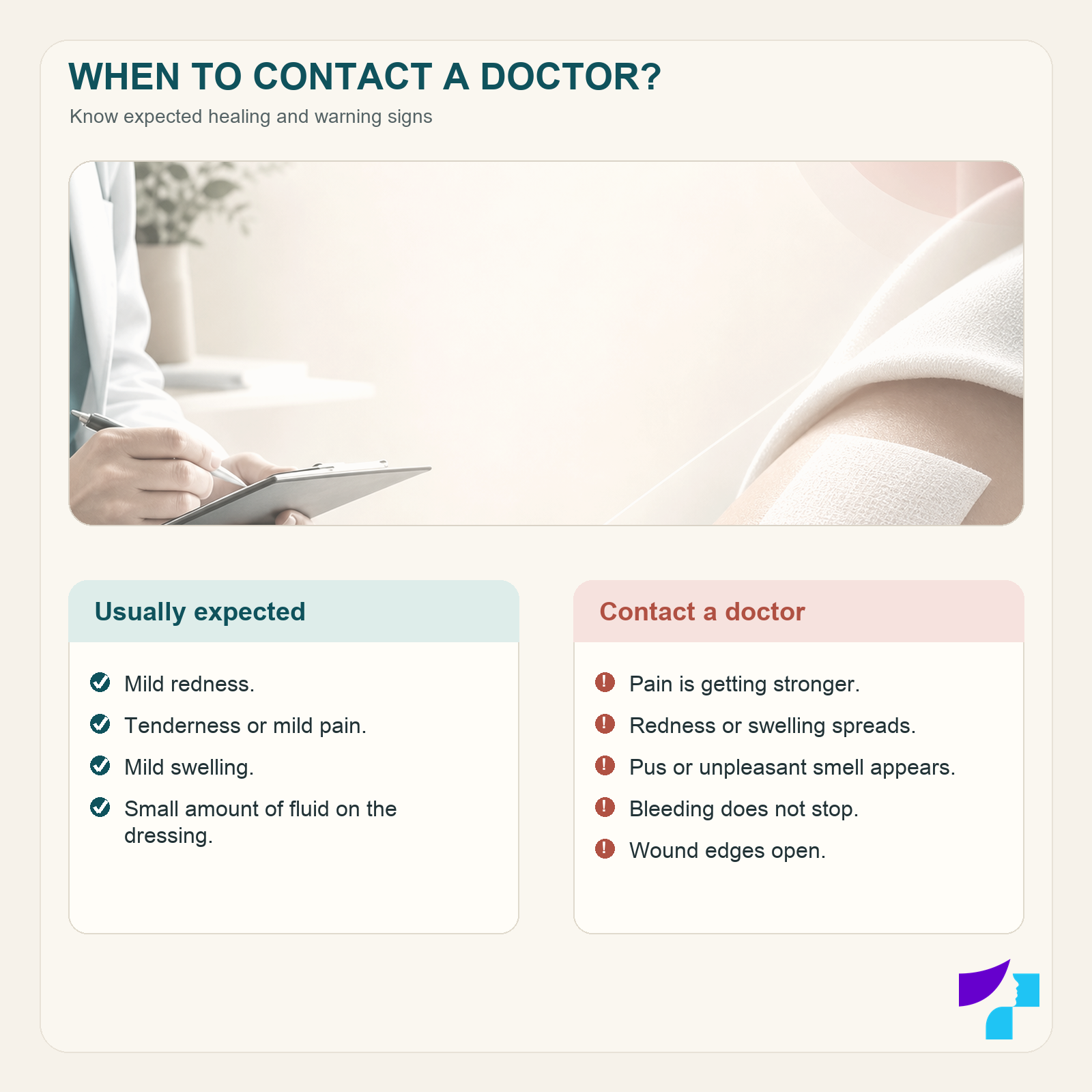
In the first days after mole removal, the following may occur:
- mild redness;
- mild swelling;
- tenderness;
- mild pain;
- a small amount of blood or clear tissue fluid on the dressing;
- itching during healing.
Other possible effects after the procedure include:
- scarring;
- lighter or darker pigmentation;
- recurrent bleeding;
- infection;
- wound edge separation;
- an allergic reaction to a dressing or care product;
- reappearance of the lesion or pigment.
When should you contact a doctor after the procedure?
A doctor or medical facility should be contacted if:
- pain becomes increasingly strong;
- redness or swelling spreads;
- the wound is noticeably hot;
- pus or unpleasant-smelling discharge appears;
- body temperature rises;
- the wound edges open;
- bleeding does not stop after prolonged direct pressure;
- a pronounced allergic reaction occurs;
- the wound does not heal for a long time;
- pigment or a nodule appears again at the removal site.
Increasing pain, heat, redness, swelling and pus-like discharge may indicate infection.
Mole removal cost
The cost of mole removal depends on the diagnosis, removal method and any additional services required.
The total cost of the visit may include:
- dermatologist consultation;
- mole examination;
- dermoscopy;
- the number of lesions to be removed;
- the size and location of the lesion;
- laser treatment or surgical excision;
- local anaesthesia;
- wound closure and dressing;
- histological examination;
- follow-up visit;
- stitch removal.
The exact procedure cost can usually be determined after the dermatologist has examined the lesion and selected the most appropriate method. The cost cannot be reliably determined from a photograph or short description alone.
Current service prices can be checked in the Medart price list or by contacting the clinic.
Mole removal in Riga
At the Medart clinic in Riga, patients can book a dermatologist consultation and mole assessment. During the visit, the doctor examines the lesion, performs dermoscopy if needed and explains whether the mole should be:
- monitored further;
- removed for medical or functional reasons;
- removed for aesthetic reasons;
- surgically excised and sent for histology.
The removal method and procedure time are determined individually. Not every mole can be removed, or is medically justified to be removed, using the method initially preferred by the patient.
Book a dermatologist consultation
Medical disclaimer
The information in this article is intended for educational and informational purposes and does not replace a dermatologist consultation, diagnosis or treatment. Moles and other skin lesions may look visually similar, but they may have different origins and require different treatment. The removal method, need for histology and aftercare are determined by the doctor after individual assessment of the patient and lesion.
References and authoritative sources
- American Academy of Dermatology — mole examination and removal
- American Academy of Dermatology — why moles and other skin lesions should not be removed at home
- American Academy of Dermatology — wound care after skin biopsy
- DermNet — melanocytic naevi, diagnostics and removal
- DermNet — melanoma diagnosis, biopsy and histological examination
- DermNet — surgical excision of skin lesions
- Guy’s and St Thomas’ NHS Foundation Trust — recovery after skin biopsy
- North Bristol NHS Trust — wound care after skin surgery
